Copyright
©The Author(s) 2022.
World J Gastroenterol. Sep 14, 2022; 28(34): 5076-5085
Published online Sep 14, 2022. doi: 10.3748/wjg.v28.i34.5076
Published online Sep 14, 2022. doi: 10.3748/wjg.v28.i34.5076
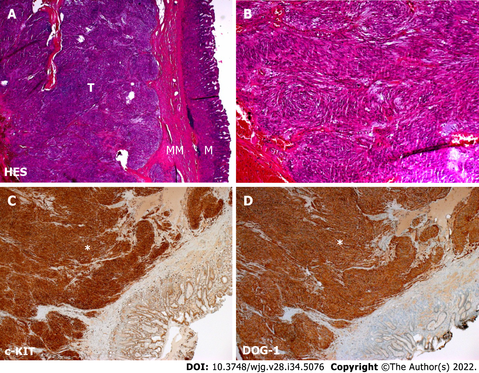
Figure 1 Histopathological features of case 1 gastrointestinal stromal tumor.
A: Hematoxylin and eosin staining showing exophytic development of a tumor from the mucosa musculus (magnification, 2.5 ×); B: Hematoxylin and eosin staining showing spindle cell morphology composed of relatively uniform cells arranged in short fascicles (magnification, 20 ×); C: Immunohistochemistry showing strong positive staining for the protooncogene c-KIT (white asterisk; magnification, 5 ×); D: Immunohistochemistry showing strong positive staining for discovered on gastrointestinal stromal tumor protein 1 (white asterisk; magnification, 5 ×). M: Mucosa; MM: Mucosa musculus; T: Tumor.
- Citation: Stammler R, Anglicheau D, Landi B, Meatchi T, Ragot E, Thervet E, Lazareth H. Gastrointestinal tumors in transplantation: Two case reports and review of literature . World J Gastroenterol 2022; 28(34): 5076-5085
- URL: https://www.wjgnet.com/1007-9327/full/v28/i34/5076.htm
- DOI: https://dx.doi.org/10.3748/wjg.v28.i34.5076


