Basic Study
Copyright ©The Author(s) 2022.
World J Gastroenterol. Sep 14, 2022; 28(34): 4993-5006
Published online Sep 14, 2022. doi: 10.3748/wjg.v28.i34.4993
Figure 1
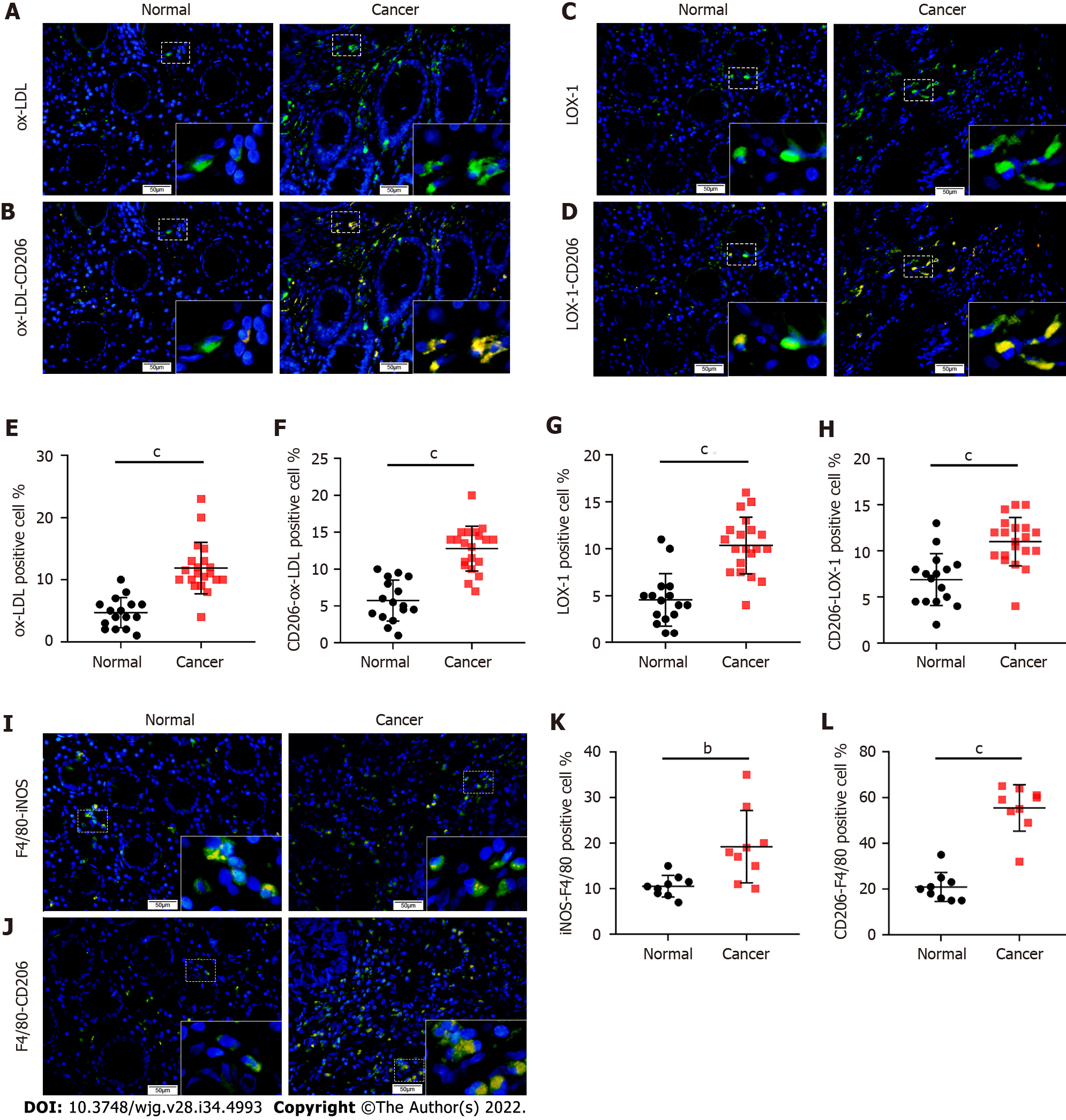
Figure 1 Increased expression of oxidized low-density lipoprotein and CD206 in the stroma of colorectal cancer tissue. A: Immunofluorescence (IF) images of oxidized low-density lipoprotein (ox-LDL) expression in colorectal tissues (scale bars represent 50 μm); B: Double IF images of CD206-ox-LDL expression in colorectal tissues (scale bars represent 50 μm); C: IF images of LOX-1 expression in colorectal tissues (scale bars represent 50 μm); D: Double IF images of CD206-LOX-1 expression in colorectal tissues (scale bars represent 50 μm); E: Histogram of ox-LDL expression in colorectal tissues based on IF results in Figure 1A; F: Quantification of CD206 and ox-LDL positive cells in colorectal tissues based on IF results in Figure 1B; G: Histogram of LOX-1 expression in colorectal tissues based on IF results in Figure 1C; H: Quantification of CD206 and LOX-1 positive cells in colorectal tissues based on IF results in Figure 1D; I: Double IF images of inducible nitric oxide synthase (iNOS)-F4/80 expression in colorectal tissues (scale bars represent 50 μm); J: Double IF images of CD206-F4/80 expression in colorectal tissues (scale bars represent 50 μm); K: Quantification of iNOS positive macrophages in colorectal tissues based on IF results in Figure 1I; L: Quantification of CD206 positive macrophages in colorectal tissues based on IF results in Figure 1J. Data are shown as the mean ± SD. Statistical analyses were conducted using an unpaired t-test. The boxed area is enlarged in the bottom right corner. bP < 0.01, cP < 0.001. ox-LDL: Oxidized low-density lipoprotein; iNOS: Inducible nitric oxide synthase.