Copyright
©The Author(s) 2022.
World J Gastroenterol. Mar 21, 2022; 28(11): 1113-1122
Published online Mar 21, 2022. doi: 10.3748/wjg.v28.i11.1113
Published online Mar 21, 2022. doi: 10.3748/wjg.v28.i11.1113
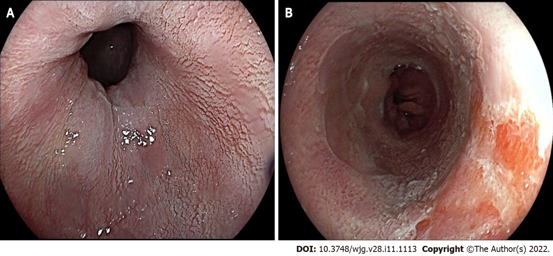
Figure 3 Acetic acid chromoendoscopy in Barrett’s esophagus.
A: Appearance of non-dysplastic mucosa (note the white, regular, and homogeneous glandular pattern); B: Early aceto-whitening loss area consistent with an intramucosal adenocarcinoma.
- Citation: Spadaccini M, Vespa E, Chandrasekar VT, Desai M, Patel HK, Maselli R, Fugazza A, Carrara S, Anderloni A, Franchellucci G, De Marco A, Hassan C, Bhandari P, Sharma P, Repici A. Advanced imaging and artificial intelligence for Barrett's esophagus: What we should and soon will do . World J Gastroenterol 2022; 28(11): 1113-1122
- URL: https://www.wjgnet.com/1007-9327/full/v28/i11/1113.htm
- DOI: https://dx.doi.org/10.3748/wjg.v28.i11.1113


