Copyright
©The Author(s) 2022.
World J Gastroenterol. Mar 14, 2022; 28(10): 1009-1023
Published online Mar 14, 2022. doi: 10.3748/wjg.v28.i10.1009
Published online Mar 14, 2022. doi: 10.3748/wjg.v28.i10.1009
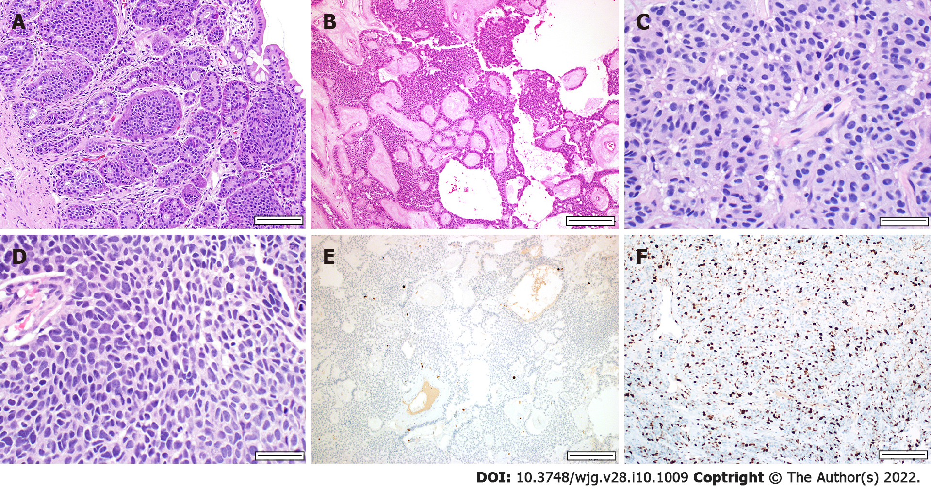
Figure 1 Morphologic features of low-grade well-differentiated neuroendocrine tumors and poorly differentiated neuroendocrine carcinoma.
A-D: Gastroenteropancreatic neuroendocrine neoplasms have a variety of architectural patterns (hematoxylin and eosin staining); A and B: Low-grade well-differentiated neuroendocrine tumors (NETs) typically have monotonous cells with round nuclei and “salt and pepper” chromatin; C: High-grade well-differentiated NETs tend to have more nuclear pleomorphism with readily identifiable mitoses; D: Small cell carcinoma, a variant of neuroendocrine carcinoma, has significant atypia with nuclear molding and scant cytoplasm. Mitoses are also readily identified; E and F: In addition to mitotic count, the Ki-67 proliferation index is necessary for grading. Low-grade well-differentiated NETs have a low proliferation index, < 20% on Ki-67 immunohistochemical (IHC) stain (E), while high-grade well-differentiated NETs have a high proliferation index, > 20% on Ki-67 IHC stain (F). Scale bars: 100 µm (A), 200 µm (B, E and F), 50 µm (C and D).
- Citation: Fang JM, Li J, Shi J. An update on the diagnosis of gastroenteropancreatic neuroendocrine neoplasms. World J Gastroenterol 2022; 28(10): 1009-1023
- URL: https://www.wjgnet.com/1007-9327/full/v28/i10/1009.htm
- DOI: https://dx.doi.org/10.3748/wjg.v28.i10.1009


