Copyright
©The Author(s) 2021.
World J Gastroenterol. Mar 7, 2021; 27(9): 794-814
Published online Mar 7, 2021. doi: 10.3748/wjg.v27.i9.794
Published online Mar 7, 2021. doi: 10.3748/wjg.v27.i9.794
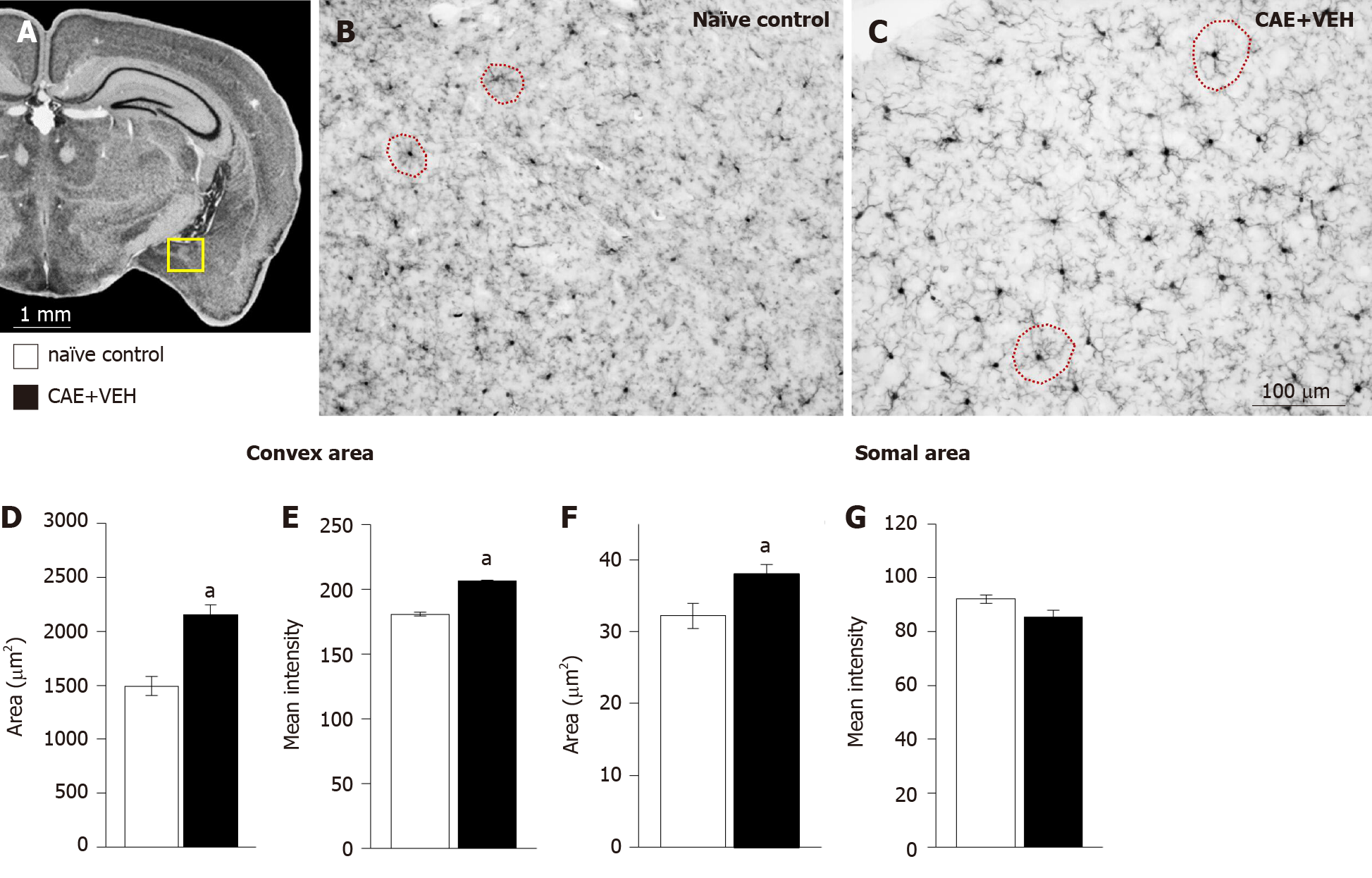
Figure 4 Microglia in the basolateral amygdala were activated only in mice with caerulein-induced pancreatitis.
A: Overview of the mouse brain shown at bregma -2.18 mm, interaural 1.62 mm[50,51]. The quantified area, the basolateral amygdala (BLA), is outlined in yellow; B and C: Examples of BLA from naïve control and vehicle treated caerulein- (CAE) induced pancreatitis mice were stained for ionized calcium-binding adaptor molecule 1; D and E: The convex area, the hull region of the microglia (samples outlined in red in B and C), was significantly enlarged and more intensely stained in mice with CAE-induced pancreatitis; F and G: Similarly, the somal area was significantly enlarged in tissue from mice with CAE-induced pancreatitis, though, the intensity of staining was not different. Naïve control n = 4, CAE + VEH n = 5, aP < 0.05 Student’s t-test. CAE: Caerulein; VEH: Vehicle.
- Citation: McIlwrath SL, Starr ME, High AE, Saito H, Westlund KN. Effect of acetyl-L-carnitine on hypersensitivity in acute recurrent caerulein-induced pancreatitis and microglial activation along the brain’s pain circuitry. World J Gastroenterol 2021; 27(9): 794-814
- URL: https://www.wjgnet.com/1007-9327/full/v27/i9/794.htm
- DOI: https://dx.doi.org/10.3748/wjg.v27.i9.794


