Copyright
©The Author(s) 2021.
World J Gastroenterol. Nov 21, 2021; 27(43): 7530-7545
Published online Nov 21, 2021. doi: 10.3748/wjg.v27.i43.7530
Published online Nov 21, 2021. doi: 10.3748/wjg.v27.i43.7530
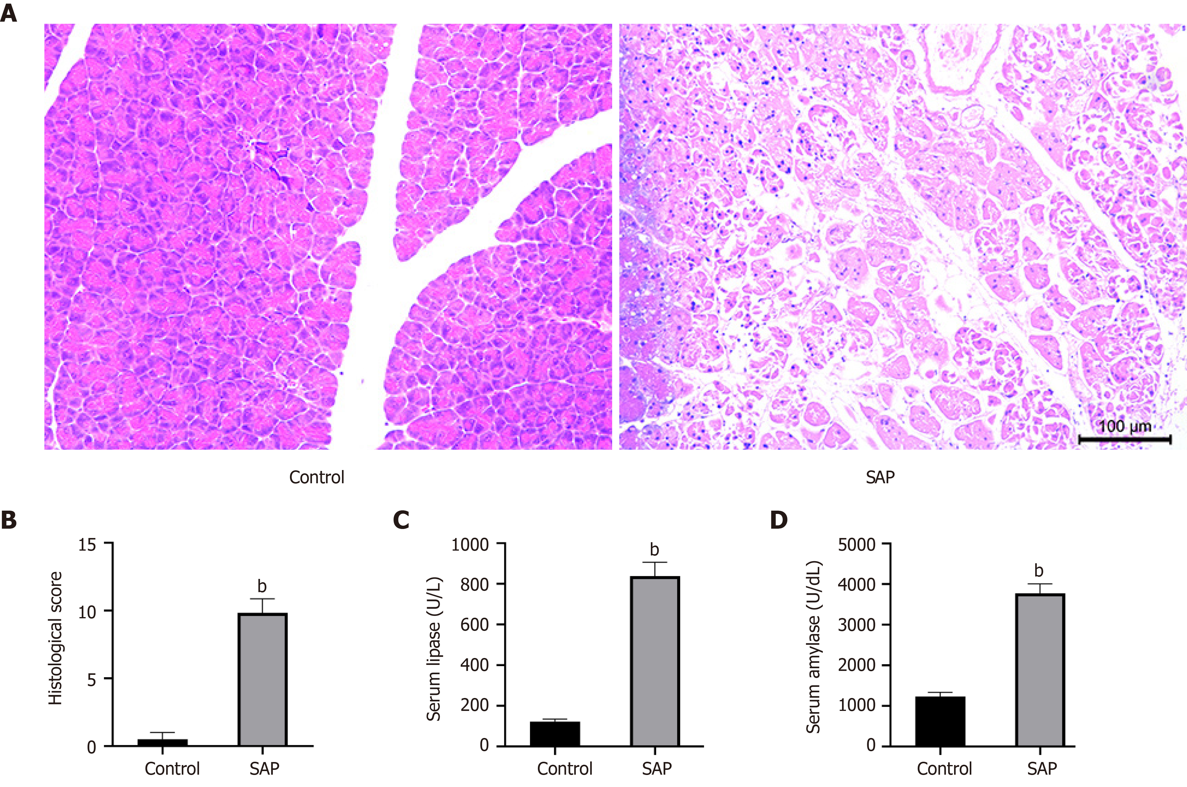
Figure 1 Evaluation of mouse model of severe acute pancreatitis.
A: Representative images of pancreatic tissues stained with hematoxylin from control (left) and severe acute pancreatitis (SAP) (right) groups (× 100 magnification); B: Histological score of pancreatic tissues in control and SAP groups; C and D: Levels of serum lipase and amylase, respectively. bP < 0.01 vs control group, n = 3 per group.
- Citation: Wu J, Yuan XH, Jiang W, Lu YC, Huang QL, Yang Y, Qie HJ, Liu JT, Sun HY, Tang LJ. Genome-wide map of N6-methyladenosine circular RNAs identified in mice model of severe acute pancreatitis. World J Gastroenterol 2021; 27(43): 7530-7545
- URL: https://www.wjgnet.com/1007-9327/full/v27/i43/7530.htm
- DOI: https://dx.doi.org/10.3748/wjg.v27.i43.7530


