Copyright
©The Author(s) 2021.
World J Gastroenterol. Sep 21, 2021; 27(35): 5908-5918
Published online Sep 21, 2021. doi: 10.3748/wjg.v27.i35.5908
Published online Sep 21, 2021. doi: 10.3748/wjg.v27.i35.5908
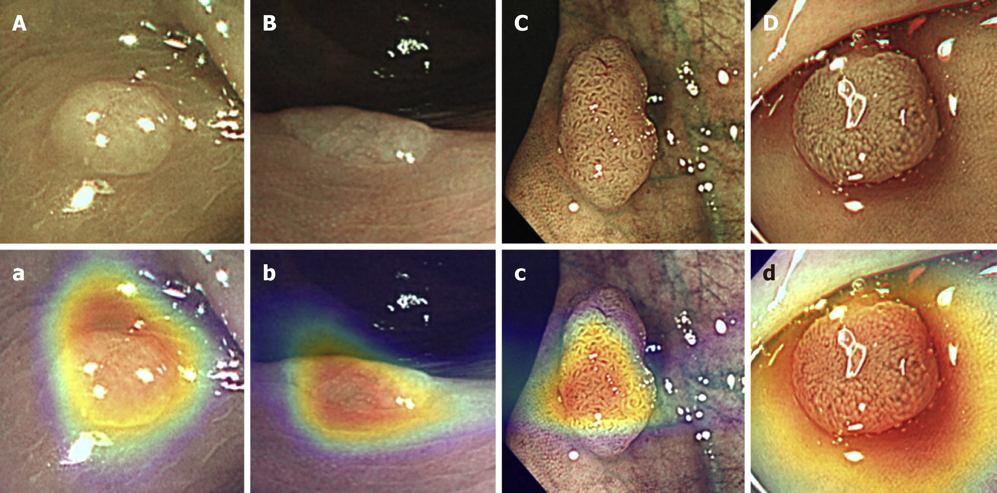
Figure 2 Illustration of coloured heatmaps, overlaid to the polyp, which demonstrates the regions that most likely contributed to the convolutional neural networks’s diagnosis.
A, B, C, D: Original narrow band imaging (NBI) of polyps; a, b, c, d: Coloured heatmap overlaid on the NBI image; Red: Higher probability that this region informed the convolutional neural networks (CNN)’s diagnosis; Blue: Lower probability that this region informed the CNN’s diagnosis. Images adapted and modified with permission from the publisher[31]. Citation: Jin EH, Lee D, Bae JH, Kang HY, Kwak MS, Seo JY, Yang JI, Yang SY, Lim SH, Yim JY, Lim JH, Chung GE, Chung SJ, Choi JM, Han YM, Kang SJ, Lee J, Chan Kim H, Kim JS. Improved Accuracy in Optical Diagnosis of Colorectal Polyps Using Convolutional Neural Networks. Gastroenterology 2020; 158(8): 2169-2179. Copyright© The Authors 2020. Published by Elsevier.
- Citation: Kader R, Hadjinicolaou AV, Georgiades F, Stoyanov D, Lovat LB. Optical diagnosis of colorectal polyps using convolutional neural networks. World J Gastroenterol 2021; 27(35): 5908-5918
- URL: https://www.wjgnet.com/1007-9327/full/v27/i35/5908.htm
- DOI: https://dx.doi.org/10.3748/wjg.v27.i35.5908


