Copyright
©The Author(s) 2021.
World J Gastroenterol. Apr 28, 2021; 27(16): 1785-1804
Published online Apr 28, 2021. doi: 10.3748/wjg.v27.i16.1785
Published online Apr 28, 2021. doi: 10.3748/wjg.v27.i16.1785
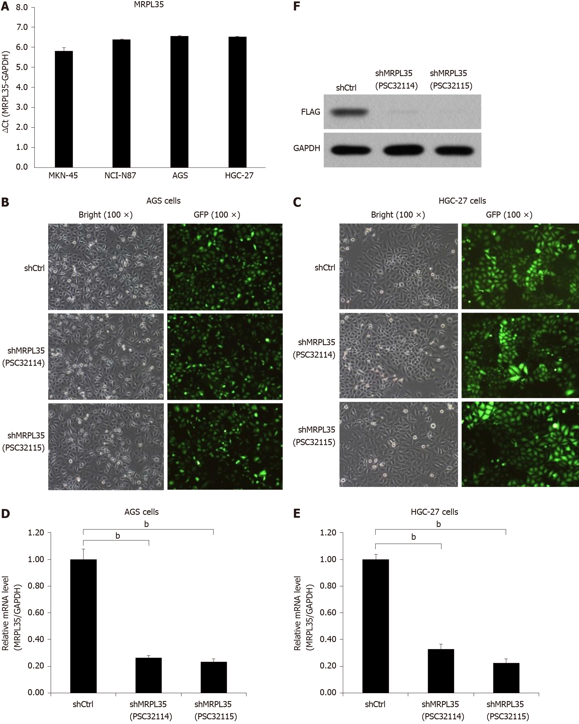
Figure 3 The expression of MRPL35 in gastric carcinoma cells.
A: Quantitative reverse transcription-polymerase chain reaction (qRT-PCR) detection of MRPL35 in gastric carcinoma cell lines (AGS, NCI-N87, MKN-45 and HGC-27); B and C: shCtrl (negative control virus) and shMRPL35 (lentiviral particles of MRPL35) (PSC32114 or PSC32115) infected AGS cells (B) and HGC-27 cells (C) at 72 h with bright and fluorescent micrographs (100 × magnification); D and E: qRT-PCR was used to analyze the expression of MRPL35 in AGS cells (D) and HGC-27 cells (E) infected with shCtrl and shMRPL35; F: Western blot was used to detect the expression of MRPL35 in AGS cells transfected with shCtrl and shMRPL35, and GAPDH (glyceraldehyde-3-phosphate dehydrogenase) was used as an internal control. n = 3. bP < 0.01. shCtrl: Negative control virus; shMRPL35: Lentiviral particles of MRPL35; GAPDH: Glyceraldehyde-3-phosphate dehydrogenase; GFP: Green fluorescent protein.
- Citation: Yuan L, Li JX, Yang Y, Chen Y, Ma TT, Liang S, Bu Y, Yu L, Nan Y. Depletion of MRPL35 inhibits gastric carcinoma cell proliferation by regulating downstream signaling proteins. World J Gastroenterol 2021; 27(16): 1785-1804
- URL: https://www.wjgnet.com/1007-9327/full/v27/i16/1785.htm
- DOI: https://dx.doi.org/10.3748/wjg.v27.i16.1785


