Copyright
©The Author(s) 2020.
World J Gastroenterol. Feb 14, 2020; 26(6): 686-695
Published online Feb 14, 2020. doi: 10.3748/wjg.v26.i6.686
Published online Feb 14, 2020. doi: 10.3748/wjg.v26.i6.686
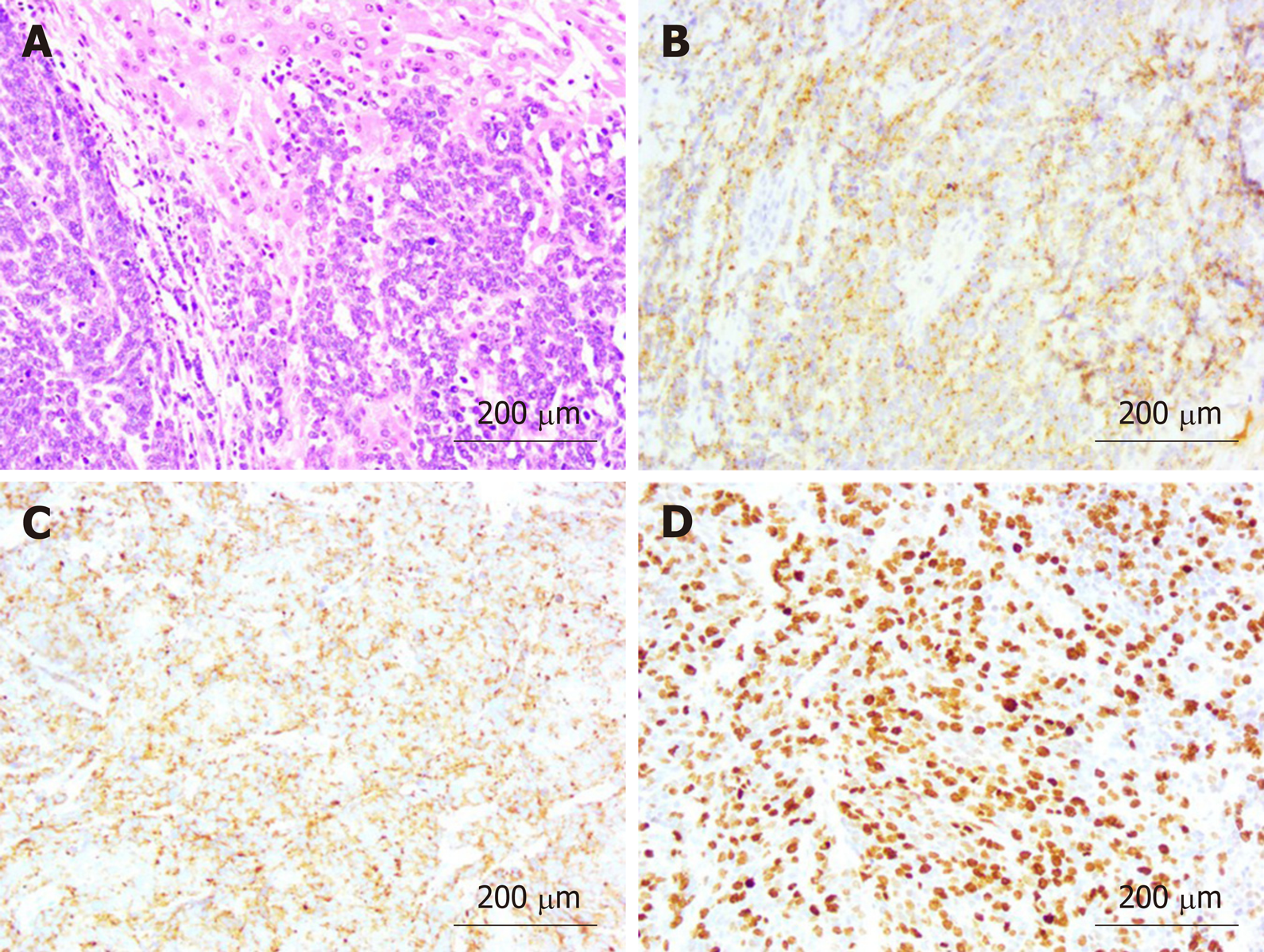
Figure 5 Pathological examination and immunohistochemical staining (20 ×).
A: Hematoxylin and eosin staining revealed poorly differentiated gallbladder neuroendocrine carcinoma with liver invasion; B: Immunohistochemical staining revealed positive expression of chromogranin A; C: Immunohistochemical staining revealed positive expression of synaptophysin; D: Immunohistochemical staining revealed that the Ki-67 index was > 80%.
- Citation: Jin M, Zhou B, Jiang XL, Zhang QY, Zheng X, Jiang YC, Yan S. Flushing as atypical initial presentation of functional gallbladder neuroendocrine carcinoma: A case report. World J Gastroenterol 2020; 26(6): 686-695
- URL: https://www.wjgnet.com/1007-9327/full/v26/i6/686.htm
- DOI: https://dx.doi.org/10.3748/wjg.v26.i6.686


