Copyright
©The Author(s) 2020.
World J Gastroenterol. Feb 14, 2020; 26(6): 686-695
Published online Feb 14, 2020. doi: 10.3748/wjg.v26.i6.686
Published online Feb 14, 2020. doi: 10.3748/wjg.v26.i6.686
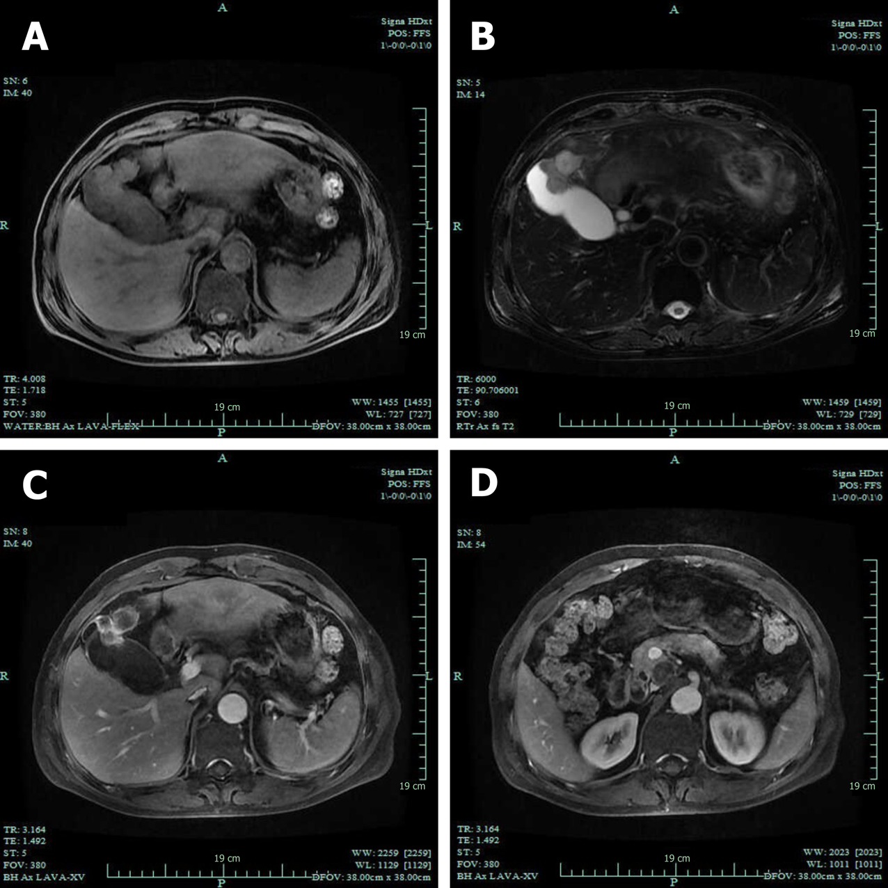
Figure 3 Magnetic resonance imaging examination.
A: The gallbladder neoplasm was revealed with liver invasion on T1-weighted imaging; B: The gallbladder neoplasm was revealed with liver invasion on T2-weighted imaging; C: The neoplasm showed increased signals in contrast-enhanced phase; D: Several enlarged lymph nodes were found in the portacaval space.
- Citation: Jin M, Zhou B, Jiang XL, Zhang QY, Zheng X, Jiang YC, Yan S. Flushing as atypical initial presentation of functional gallbladder neuroendocrine carcinoma: A case report. World J Gastroenterol 2020; 26(6): 686-695
- URL: https://www.wjgnet.com/1007-9327/full/v26/i6/686.htm
- DOI: https://dx.doi.org/10.3748/wjg.v26.i6.686


