Copyright
©The Author(s) 2020.
World J Gastroenterol. Dec 21, 2020; 26(47): 7584-7592
Published online Dec 21, 2020. doi: 10.3748/wjg.v26.i47.7584
Published online Dec 21, 2020. doi: 10.3748/wjg.v26.i47.7584
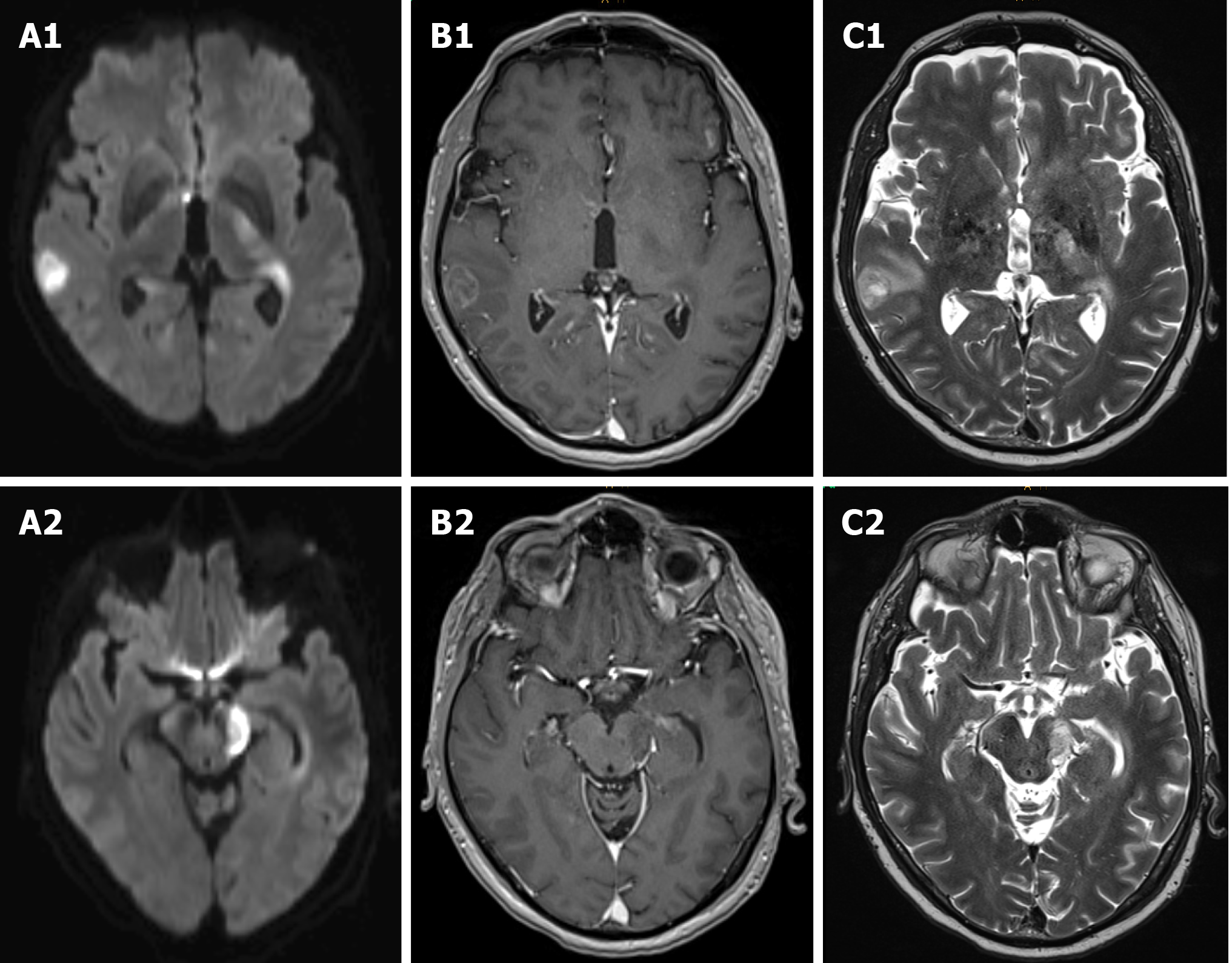
Figure 3 Cerebral magnetic resonance imaging images.
Cerebral magnetic resonance imaging demonstrated multifocal lesions in right temporal lobe, periventricular third ventricle right and lateral ventricle left, basal ganglia left and mesencephalon left with diffusion restriction on diffusion-weighted images as well as inhomogeneous and circular contrast enhancement as well as hyperintensities on T2-weighted images. A: Diffusion-weighted images (A1 and 2); B: Inhomogeneous and circular contrast enhancement (B1 and 2); C: T2-weighted images (C1 and 2).
- Citation: Horvath L, Oberhuber G, Chott A, Effenberger M, Tilg H, Gunsilius E, Wolf D, Iglseder S. Multiple cerebral lesions in a patient with refractory celiac disease: A case report. World J Gastroenterol 2020; 26(47): 7584-7592
- URL: https://www.wjgnet.com/1007-9327/full/v26/i47/7584.htm
- DOI: https://dx.doi.org/10.3748/wjg.v26.i47.7584


