Copyright
©The Author(s) 2020.
World J Gastroenterol. Jan 28, 2020; 26(4): 448-455
Published online Jan 28, 2020. doi: 10.3748/wjg.v26.i4.448
Published online Jan 28, 2020. doi: 10.3748/wjg.v26.i4.448
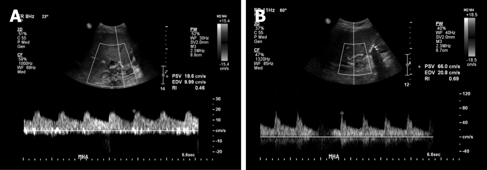
Figure 2 Images of doppler ultrasound in the main hepatic artery.
A: Doppler ultrasound in the main hepatic artery prior to intervention (day 80 post-transplant) demonstrating parvus et tardus waveform with low resistive index and low peak systolic velocity; B: After stenting (day 115 post-transplant, day 15 post stenting) showing normal waveform, normal peak systolic velocity, and normal resistive index.
- Citation: Barahman M, Alanis L, DiNorcia J, Moriarty JM, McWilliams JP. Hepatic artery stenosis angioplasty and implantation of Wingspan neurovascular stent: A case report and discussion of stenting in tortuous vessels. World J Gastroenterol 2020; 26(4): 448-455
- URL: https://www.wjgnet.com/1007-9327/full/v26/i4/448.htm
- DOI: https://dx.doi.org/10.3748/wjg.v26.i4.448


