Copyright
©The Author(s) 2020.
World J Gastroenterol. Aug 14, 2020; 26(30): 4557-4563
Published online Aug 14, 2020. doi: 10.3748/wjg.v26.i30.4557
Published online Aug 14, 2020. doi: 10.3748/wjg.v26.i30.4557
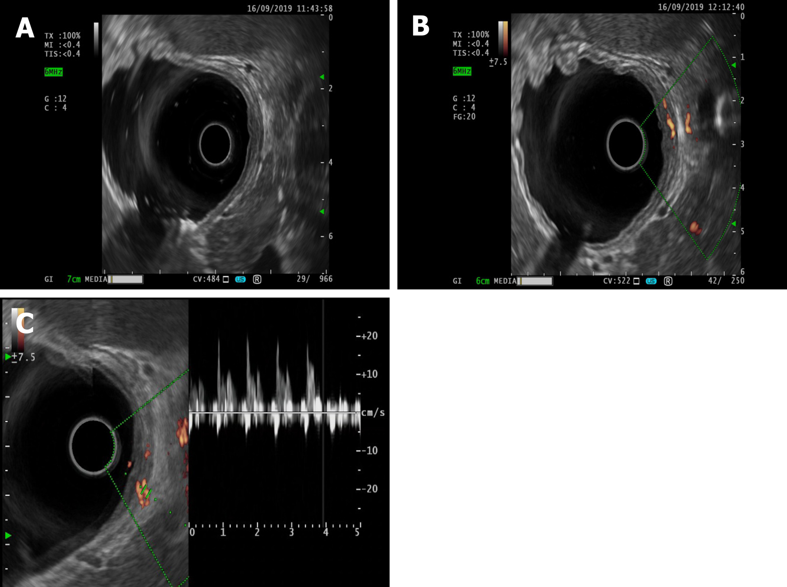
Figure 3 Endoscopic ultrasonography.
A: Dieulafoy’s lesion under endoscopic ultrasonography; B: Ultrasonic Doppler showing the blood flow signal of the mycomembrane muscle and submucosa in the lesion area; C: Doppler spectrum representing the arterial blood flow spectrum.
- Citation: Yu S, Wang XM, Chen X, Xu HY, Wang GJ, Ni N, Sun YX. Endoscopic full-thickness resection to treat active Dieulafoy's disease: A case report. World J Gastroenterol 2020; 26(30): 4557-4563
- URL: https://www.wjgnet.com/1007-9327/full/v26/i30/4557.htm
- DOI: https://dx.doi.org/10.3748/wjg.v26.i30.4557


