Copyright
©The Author(s) 2020.
World J Gastroenterol. Jul 21, 2020; 26(27): 3899-3916
Published online Jul 21, 2020. doi: 10.3748/wjg.v26.i27.3899
Published online Jul 21, 2020. doi: 10.3748/wjg.v26.i27.3899
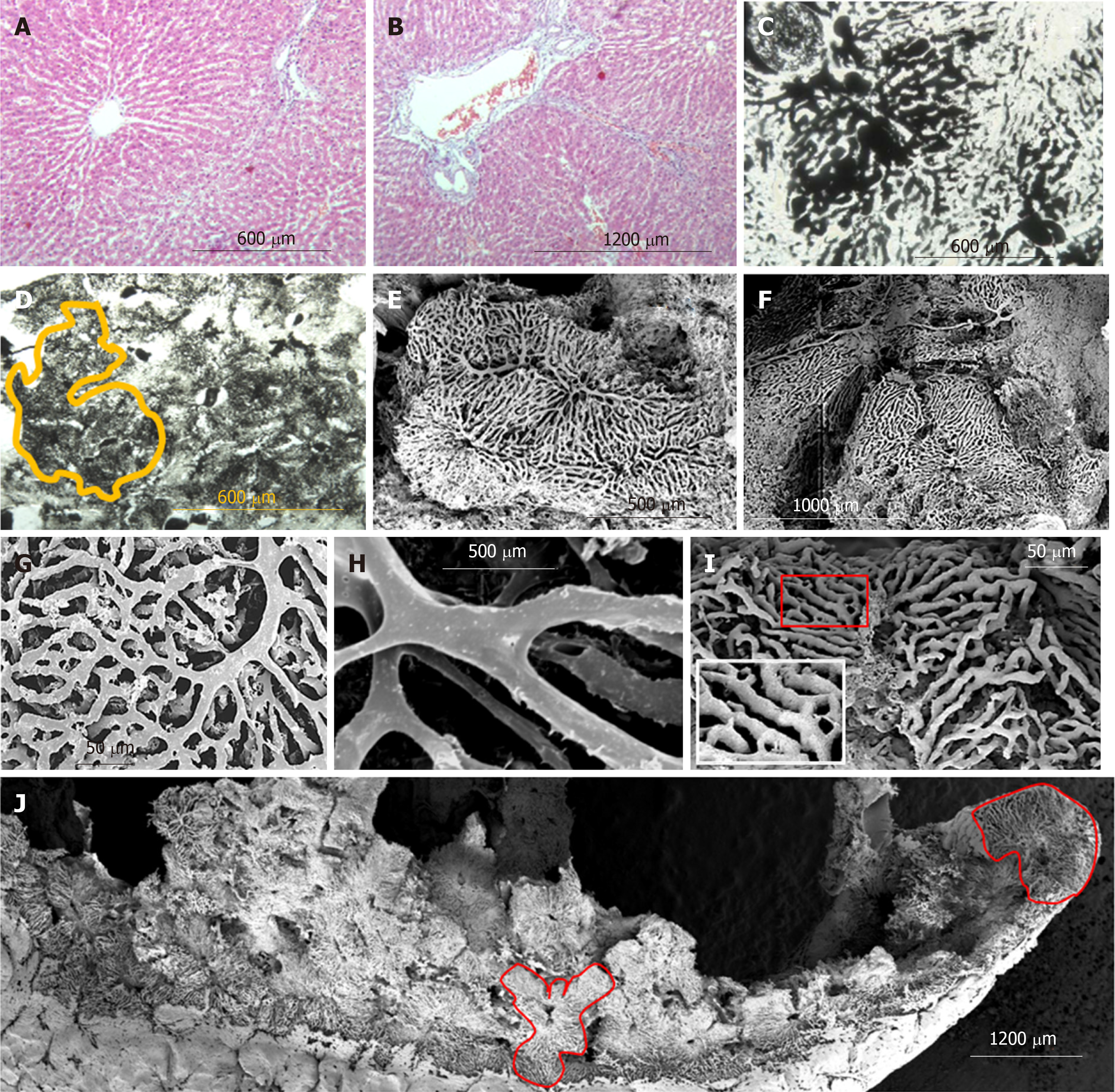
Figure 4 Histology and scanning electron microscopy of corrosion casts of regenerated liver (SG1).
A and B: Liver tissue histology (Masson’s Trichrome); C: Liver lobules (histology after Indian ink – gelatin injection); D: “mega-lobule” formed by adjacent lobules with intercommunicated sinusoidal meshwork (bordered by yellow line) (histology after Indian ink – gelatin injection); E-J: Scanning electron microscopy (SEM) of corrosion casts of liver vessels; E and F: “mega-lobules”; G-I: SEM of corrosion casts of sinusoids; J: SEM of vascular corrosion casts of liver lobe; “mega-lobules” (bordered by red line).
- Citation: Tsomaia K, Patarashvili L, Karumidze N, Bebiashvili I, Azmaipharashvili E, Modebadze I, Dzidziguri D, Sareli M, Gusev S, Kordzaia D. Liver structural transformation after partial hepatectomy and repeated partial hepatectomy in rats: A renewed view on liver regeneration. World J Gastroenterol 2020; 26(27): 3899-3916
- URL: https://www.wjgnet.com/1007-9327/full/v26/i27/3899.htm
- DOI: https://dx.doi.org/10.3748/wjg.v26.i27.3899


