Copyright
©The Author(s) 2020.
World J Gastroenterol. Jun 14, 2020; 26(22): 3110-3117
Published online Jun 14, 2020. doi: 10.3748/wjg.v26.i22.3110
Published online Jun 14, 2020. doi: 10.3748/wjg.v26.i22.3110
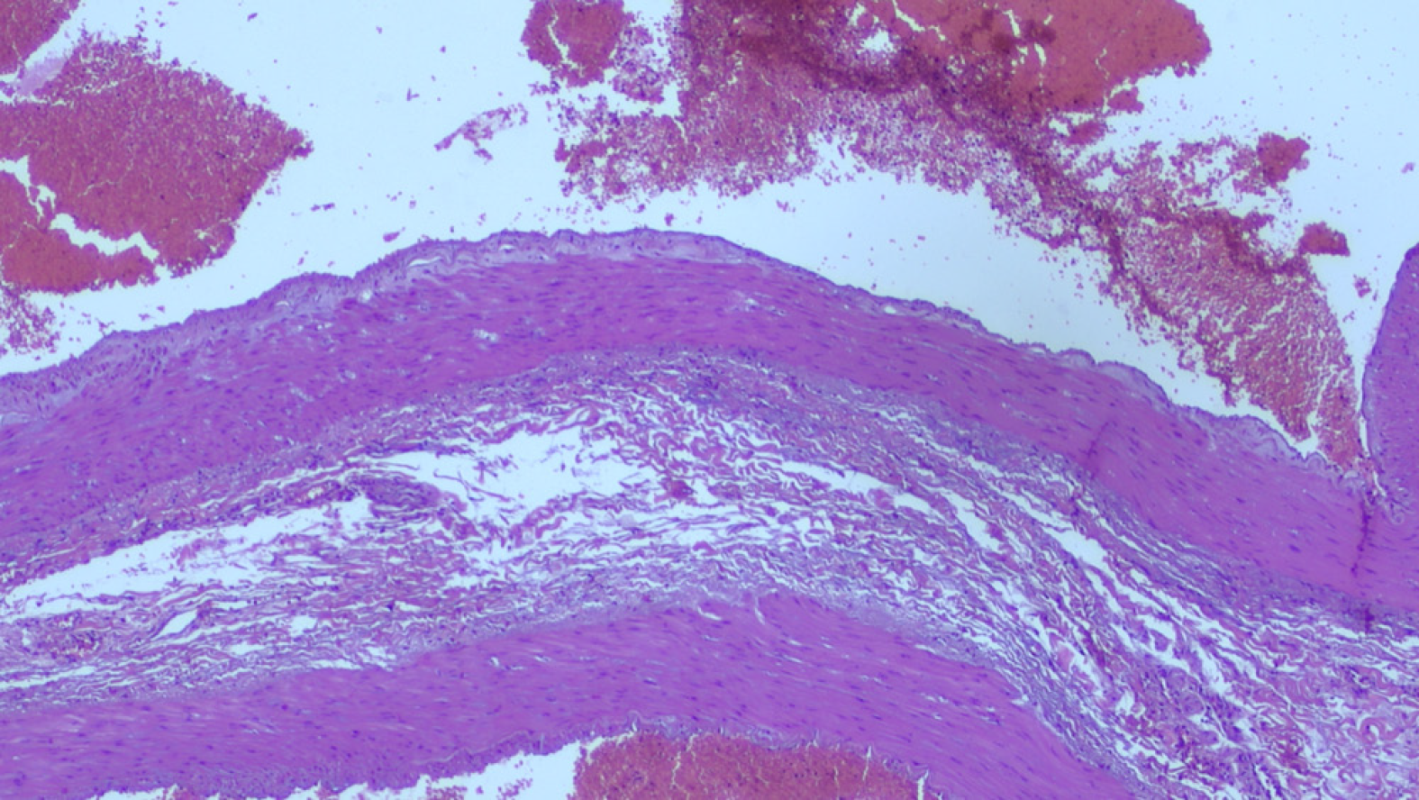
Figure 3 Morphological findings in keeping with aneurysm of the splenic artery.
At histology (surgical specimen stained with haematoxylin and eosin at 4 ×) there is evidence of marked alterations of the vascular wall, with destruction of the internal elastic lamina and partial dissection, myxoid degeneration and phlogistic infiltrate, smooth muscle hyperplasia in tunica media, destruction of the elastic fibers of the vessel wall.
- Citation: Panzera F, Inchingolo R, Rizzi M, Biscaglia A, Schievenin MG, Tallarico E, Pacifico G, Di Venere B. Giant splenic artery aneurysm presenting with massive upper gastrointestinal bleeding: A case report and review of literature. World J Gastroenterol 2020; 26(22): 3110-3117
- URL: https://www.wjgnet.com/1007-9327/full/v26/i22/3110.htm
- DOI: https://dx.doi.org/10.3748/wjg.v26.i22.3110


