Copyright
©The Author(s) 2020.
World J Gastroenterol. Jun 7, 2020; 26(21): 2831-2838
Published online Jun 7, 2020. doi: 10.3748/wjg.v26.i21.2831
Published online Jun 7, 2020. doi: 10.3748/wjg.v26.i21.2831
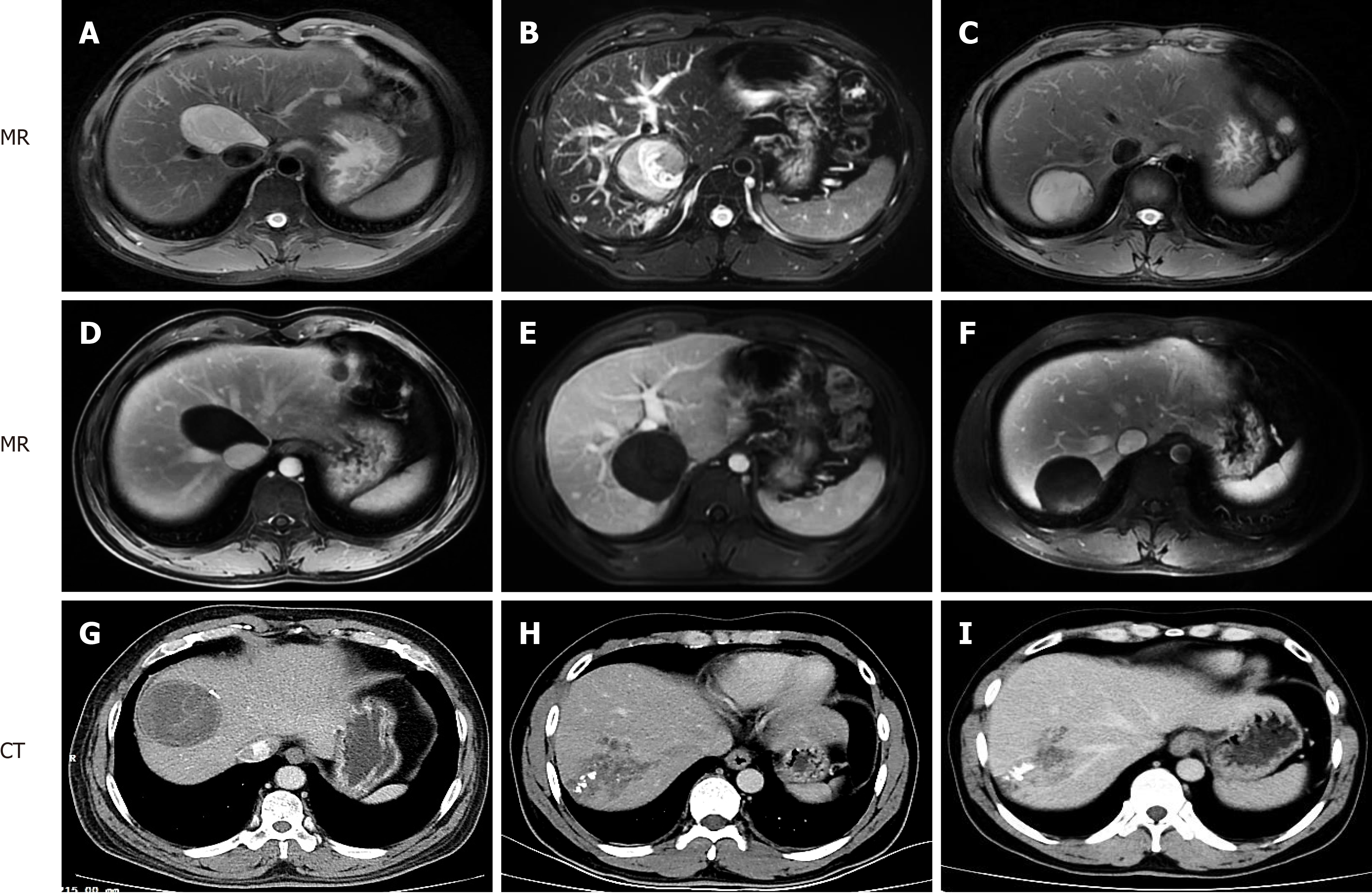
Figure 1 Contrast-enhanced magnetic resonance imaging and computed tomography manifestations of hepatic cystic and alveolar echinococcosis.
A, D: Patient 1, cystic echinococcosis in caudate lobe; B, E: Patient 2, cystic echinococcosis in caudate lobe; C, F: Patient 3, cystic echinococcosis in segment VII; G: Patient 4, cystic echinococcosis in segment VIII; H, I: Patient 5, alveolar echinococcosis in segment VII/VIII. MR: Magnetic resonance; CT: Computed tomography.
- Citation: Zhao ZM, Yin ZZ, Meng Y, Jiang N, Ma ZG, Pan LC, Tan XL, Chen X, Liu R. Successful robotic radical resection of hepatic echinococcosis located in posterosuperior liver segments. World J Gastroenterol 2020; 26(21): 2831-2838
- URL: https://www.wjgnet.com/1007-9327/full/v26/i21/2831.htm
- DOI: https://dx.doi.org/10.3748/wjg.v26.i21.2831


