Copyright
©The Author(s) 2018.
World J Gastroenterol. Mar 7, 2018; 24(9): 1056-1062
Published online Mar 7, 2018. doi: 10.3748/wjg.v24.i9.1056
Published online Mar 7, 2018. doi: 10.3748/wjg.v24.i9.1056
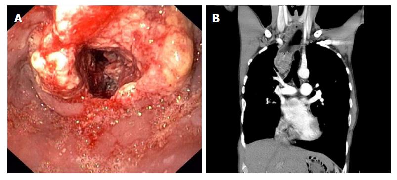
Figure 2 Findings at upper endoscopy and chest computed tomography scan (CT scan) (case 2).
A: Upper endoscopy revealing a stenotic ulcerative tumor in the proximal esophagus, 22-29 cm from incisors. Histological examination of esophageal biopsies confirmed the diagnosis esophageal squamous cell carcinoma. B: Chest CT scan showing a tumor mass in the proximal esophagus with suspected tumor invasion in the trachea.
- Citation: Vergouwe FW, Gottrand M, Wijnhoven BP, IJsselstijn H, Piessen G, Bruno MJ, Wijnen RM, Spaander MC. Four cancer cases after esophageal atresia repair: Time to start screening the upper gastrointestinal tract. World J Gastroenterol 2018; 24(9): 1056-1062
- URL: https://www.wjgnet.com/1007-9327/full/v24/i9/1056.htm
- DOI: https://dx.doi.org/10.3748/wjg.v24.i9.1056


