Copyright
©The Author(s) 2018.
World J Gastroenterol. Jan 14, 2018; 24(2): 297-302
Published online Jan 14, 2018. doi: 10.3748/wjg.v24.i2.297
Published online Jan 14, 2018. doi: 10.3748/wjg.v24.i2.297
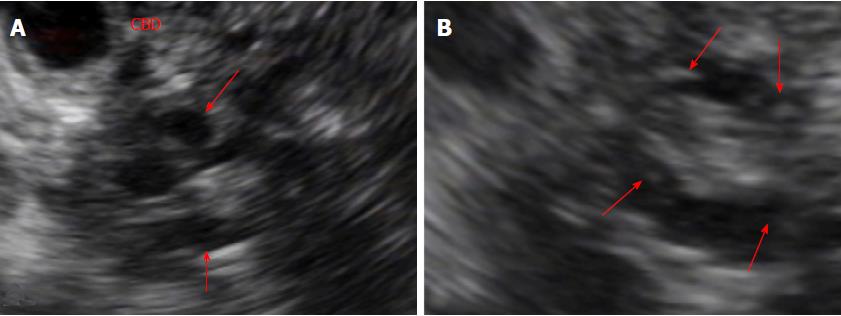
Figure 3 Findings from endoscopic ultrasound.
A: EUS shows a few anechoic tubular structures (arrows), causing indentation of distal CBD and dilatation of proximal bile duct; B: EUS shows small hyperechoic mural nodules (arrows) in the dilated branch pancreatic ducts. EUS: Endoscopic ultrasound; CBD: Common bile duct.
- Citation: Jee KN. Mass forming chronic pancreatitis mimicking pancreatic cystic neoplasm: A case report. World J Gastroenterol 2018; 24(2): 297-302
- URL: https://www.wjgnet.com/1007-9327/full/v24/i2/297.htm
- DOI: https://dx.doi.org/10.3748/wjg.v24.i2.297


