Copyright
©The Author(s) 2017.
World J Gastroenterol. Feb 14, 2017; 23(6): 986-998
Published online Feb 14, 2017. doi: 10.3748/wjg.v23.i6.986
Published online Feb 14, 2017. doi: 10.3748/wjg.v23.i6.986
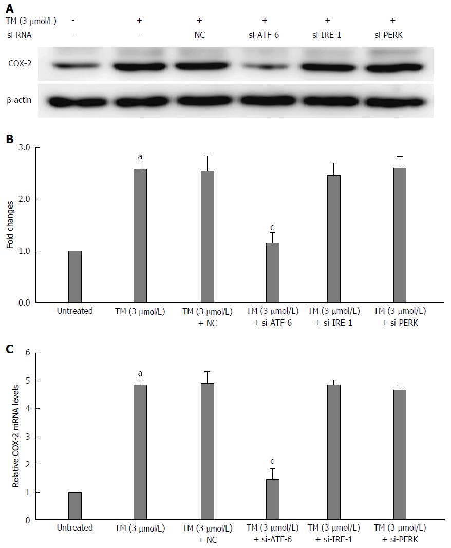
Figure 4 Relationship between activating transcription factor 6 and cyclooxygenase-2 under endoplasmic reticulum stress.
The mRNA of the unfolded protein response (UPR) pathways was interfered by ATF-6, IRE-1 and PERK siRNA after pretreatment by tunicamycin (TM) for 8 h. A: Equal protein amounts of cell lysates were subjected to western blot assay using anti-COX-2. β-actin in the same HepG2 cell extract was used as an internal reference. B: Optical density reading values of specific proteins are represented as fold-differences relative to the loading control protein, β-actin. aP < 0.01 vs the negative control. RNA was harvested and gene expression examined by qRT-PCR in the same condition above. C: The qRT-PCR fold-changes were normalized using the expression of housekeeping gene (GAPDH) and vs those obtained from untreated HepG2 cells. aP < 0.01 vs the negative control; cP < 0.01 vs the untreated HepG2 cells. ATF-6: Activating transcription factor 6; NC: Negative control; COX-2: Cyclooxygenase-2; IRE: Inositol-requiring enzyme; PERK: Protein kinase RNA-like endoplasmic reticulum kinase.
- Citation: Bu LJ, Yu HQ, Fan LL, Li XQ, Wang F, Liu JT, Zhong F, Zhang CJ, Wei W, Wang H, Sun GP. Melatonin, a novel selective ATF-6 inhibitor, induces human hepatoma cell apoptosis through COX-2 downregulation. World J Gastroenterol 2017; 23(6): 986-998
- URL: https://www.wjgnet.com/1007-9327/full/v23/i6/986.htm
- DOI: https://dx.doi.org/10.3748/wjg.v23.i6.986


