Copyright
©The Author(s) 2017.
World J Gastroenterol. Dec 21, 2017; 23(47): 8367-8375
Published online Dec 21, 2017. doi: 10.3748/wjg.v23.i47.8367
Published online Dec 21, 2017. doi: 10.3748/wjg.v23.i47.8367
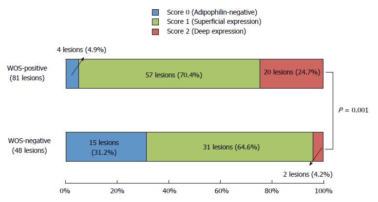
Figure 3 Comparison of the distribution of adipophilin between white opaque substance-positive and -negative lesions.
The scores for adipophilin expression depth are significantly different between the two groups of lesions (P = 0.001), with a predominance of score 2 (deep expression) in WOS-positive lesions (24.7%) compared to WOS-negative lesions (4.2%).
- Citation: Kawasaki K, Eizuka M, Nakamura S, Endo M, Yanai S, Akasaka R, Toya Y, Fujita Y, Uesugi N, Ishida K, Sugai T, Matsumoto T. Association between white opaque substance under magnifying colonoscopy and lipid droplets in colorectal epithelial neoplasms. World J Gastroenterol 2017; 23(47): 8367-8375
- URL: https://www.wjgnet.com/1007-9327/full/v23/i47/8367.htm
- DOI: https://dx.doi.org/10.3748/wjg.v23.i47.8367


