Copyright
©The Author(s) 2017.
World J Gastroenterol. Nov 14, 2017; 23(42): 7505-7518
Published online Nov 14, 2017. doi: 10.3748/wjg.v23.i42.7505
Published online Nov 14, 2017. doi: 10.3748/wjg.v23.i42.7505
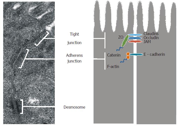
Figure 2 Ultrastructure and corresponding schematic representation of intercellular junctions.
Transmission electron microscopy (JEOL JEM-1011, Japan; × 60000) was used to show the ultrastructure of intercellular junctions in the human small intestine. The transmission electron micrograph comes from our own research. JAM: Junction adhesion molecule.
- Citation: Cukrowska B, Sowińska A, Bierła JB, Czarnowska E, Rybak A, Grzybowska-Chlebowczyk U. Intestinal epithelium, intraepithelial lymphocytes and the gut microbiota - Key players in the pathogenesis of celiac disease. World J Gastroenterol 2017; 23(42): 7505-7518
- URL: https://www.wjgnet.com/1007-9327/full/v23/i42/7505.htm
- DOI: https://dx.doi.org/10.3748/wjg.v23.i42.7505


