Copyright
©The Author(s) 2017.
World J Gastroenterol. Jun 14, 2017; 23(22): 4016-4038
Published online Jun 14, 2017. doi: 10.3748/wjg.v23.i22.4016
Published online Jun 14, 2017. doi: 10.3748/wjg.v23.i22.4016
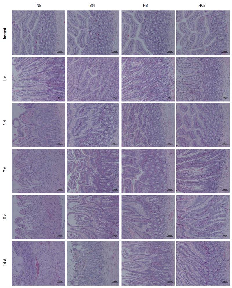
Figure 4 Pathology of transplanted small bowel (HE staining, × 100).
All four groups showed normal histological results immediately after transplant. The NS group showed mild rejection at day 1 after transplantation: shorter and bifurcated intestinal villi, mild submucosal edema, cryptic epithelial cells with mild damage (cytoplasmic basophilic increase, nuclear enlargement, and color deepening) and increased apoptosis, more than six apoptotic bodies per 10 crypt cells, and mild mononuclear cells which are the main inflammatory cells found during inflammatory infiltration of the lamina propria; in contrast, the BM group, HB group, and HCB group showed only mild edema. The pathological changes at 3 d after transplantation in the NS group were more severe than those at 1 day, where the submucosal edema was aggravated, and there was increased inflammatory infiltration of the lamina propria; the BM group and HB group showed mild inflammatory infiltration of the lamina propria, and the HCB group was similar to the normal intestine. The NS group showed moderate rejection at day 7 after transplantation: a reduced ratio of villous height to crypt, partial necrosis of glandular epithelial cells, aggravated edema and inflammation, diffuse crypt damage and increased apoptosis, and mild arteritis and congestion of lamina propria and submucosa; the BM group and HB group showed mild rejection: the BM group had shorter intestinal villi, mild submucosal edema, inflammatory cell infiltration, mild crypt epithelial injury, and increased apoptosis; the HB group had mild inflammatory cell infiltration, mild crypt injury, with other signs not being obvious; the HCB group were indeterminate for rejection: mild cryptic epithelial damage and the number of apoptotic bodies increased, with mild, local inflammatory cell infiltration of the lamina propria. The NS group showed severe rejection at day 10 after transplantation: intestinal villus changes were further aggravated, with serious shedding of the intestinal mucosal epithelial cells, while crypt epithelial injury was very serious; inflammatory cell infiltration involved the muscle layer, resulting in severe arteritis; the pathological changes in the BM group, HB group, and HCB group at day 10 were more severe than those at day 7 after transplantation. The structures of the intestinal mucosa layer were completely destroyed and the intestinal wall became thinner with necrosis in the NS group 14 d after transplantation; the BM group showed moderate rejection; the HB group did not achieve moderate rejection but rejection was more severe than mild; the HCB group showed mild rejection.
- Citation: Yin ML, Song HL, Yang Y, Zheng WP, Liu T, Shen ZY. Effect of CXCR3/HO-1 genes modified bone marrow mesenchymal stem cells on small bowel transplant rejection. World J Gastroenterol 2017; 23(22): 4016-4038
- URL: https://www.wjgnet.com/1007-9327/full/v23/i22/4016.htm
- DOI: https://dx.doi.org/10.3748/wjg.v23.i22.4016


