Copyright
©The Author(s) 2017.
World J Gastroenterol. Jan 14, 2017; 23(2): 256-264
Published online Jan 14, 2017. doi: 10.3748/wjg.v23.i2.256
Published online Jan 14, 2017. doi: 10.3748/wjg.v23.i2.256
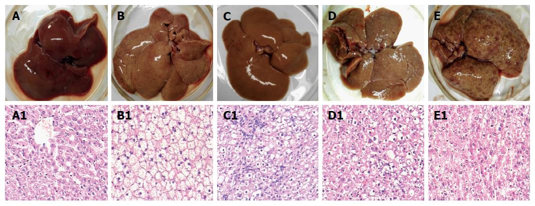
Figure 1 Rat liver tissues and their pathological examination.
Liver alterations after the rats were sacrificed at different stages during hepatocyte malignant transformation. A: A representative specimen from the control rats with normal diet; B: A representative specimen from the rats with high-fat diet without 2-fluorenyl acetamide (2-FAA); C: A representative specimen from the rats with high-fat diet containing 2-FAA, at early stage; D: A representative specimen from the rats with high-fat diet containing 2-FAA, at interim stage; and E: A representative specimen from the rats with high-fat diet containing 2-FAA, at later stage. The liver sections were examined with hematoxylin and eosin staining and then divided into the control (A1), fatty liver (B1), degeneration (C1), precancerous (D1), and cancerous (E1) groups; A1-E1: The original magnification of the corresponding rat liver sections was × 200.
- Citation: Gu JJ, Yao M, Yang J, Cai Y, Zheng WJ, Wang L, Yao DB, Yao DF. Mitochondrial carnitine palmitoyl transferase-II inactivity aggravates lipid accumulation in rat hepatocarcinogenesis. World J Gastroenterol 2017; 23(2): 256-264
- URL: https://www.wjgnet.com/1007-9327/full/v23/i2/256.htm
- DOI: https://dx.doi.org/10.3748/wjg.v23.i2.256


