Copyright
©The Author(s) 2017.
World J Gastroenterol. May 21, 2017; 23(19): 3407-3417
Published online May 21, 2017. doi: 10.3748/wjg.v23.i19.3407
Published online May 21, 2017. doi: 10.3748/wjg.v23.i19.3407
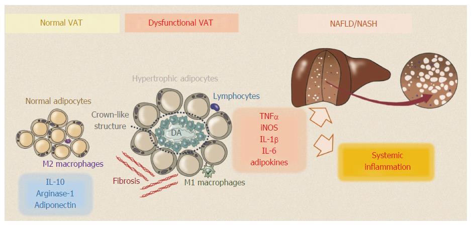
Figure 1 Normal visceral adipose tissue consists of a loose connective tissue that is populated with tightly packed adipocytes.
In lean individuals VAT homeostasis is maintained by adiponectin released by adipocytes and by M2 macrophages through the secretion of anti-inflammatory cytokines, such as interleukin (IL)-10 and arginase-1. During obesity, dysfunctional visceral adipose tissue (VAT) undergoes excessive fibrosis and accumulation of inflammatory cells. Active macrophages surround dying adipocytes (DA) in typical “crown-like structures”. Pro-M1 polarized macrophages secrete pro-inflammatory cytokines including TNFα, IL-1β and IL-6, which can promote chronic local and systemic inflammation. VAT secretes a large number of adipokines which could play a pivotal role in development of NAFLD. NAFLD: Non-alcoholic fatty liver disease.
- Citation: Cimini FA, Barchetta I, Carotti S, Bertoccini L, Baroni MG, Vespasiani-Gentilucci U, Cavallo MG, Morini S. Relationship between adipose tissue dysfunction, vitamin D deficiency and the pathogenesis of non-alcoholic fatty liver disease. World J Gastroenterol 2017; 23(19): 3407-3417
- URL: https://www.wjgnet.com/1007-9327/full/v23/i19/3407.htm
- DOI: https://dx.doi.org/10.3748/wjg.v23.i19.3407


