Copyright
©The Author(s) 2017.
World J Gastroenterol. Apr 7, 2017; 23(13): 2443-2447
Published online Apr 7, 2017. doi: 10.3748/wjg.v23.i13.2443
Published online Apr 7, 2017. doi: 10.3748/wjg.v23.i13.2443
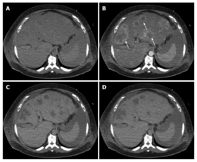
Figure 2 Multi-phase axial computerized tomography images of hepatic angiosarcoma.
A: Precontrast image fails to visualize the tumor likely due to its isodensity; B: Arterial phase demonstrates discrete, multifocal masses with peripheral enhancement (arrow); C and D: Portal venous and delayed phase images show discrete masses without enhancement and without definite washout.
- Citation: Wadhwa S, Kim TH, Lin L, Kanel G, Saito T. Hepatic angiosarcoma with clinical and histological features of Kasabach-Merritt syndrome. World J Gastroenterol 2017; 23(13): 2443-2447
- URL: https://www.wjgnet.com/1007-9327/full/v23/i13/2443.htm
- DOI: https://dx.doi.org/10.3748/wjg.v23.i13.2443


