Copyright
©The Author(s) 2017.
World J Gastroenterol. Mar 21, 2017; 23(11): 2090-2094
Published online Mar 21, 2017. doi: 10.3748/wjg.v23.i11.2090
Published online Mar 21, 2017. doi: 10.3748/wjg.v23.i11.2090
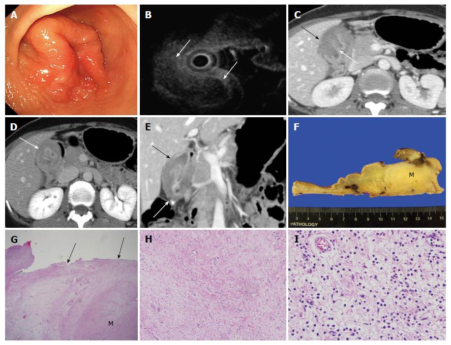
Figure 1 A 43-year-old female with an inflammatory fibroid polyp with massive fibrosis.
A: Endoscopy showed an approximately 4 cm mass-like lesion with mucosal edema and superficial ulcer on the gastric antrum; B: On endoscopic ultrasound examination, an approximately 4 cm heterogeneous hypoechoic submucosal mass-like lesion (arrows) was seen; C: The portal phase of axial contrast-enhanced computed tomography (CT) scan showed a hypoattenuated marked wall thickening of the submucosal layer at the gastric antrum (black arrow) with preserved mucosal enhancement (white arrow); D: The 3-min delayed phase of axial contrast-enhanced CT scan demonstrated delayed enhancement (about 115 HU) of the submucosal layer at the gastric antrum (arrow); E: The coronal image of contrast-enhanced CT scan revealed that the lesion did not extend to the liver and demonstrated no perigastric fat infiltration (arrows); F: The surgical specimen demonstrated a submucosal tumor (M) measuring 4.5 cm × 4.0 cm × 3.0 cm and the overlying mucosa was intact; G: Microscopic examination (hematoxylin and eosin stain, magnification × 12.5) demonstrated a submucosal mass-like lesion (M). The borders of the submucosal mass-like lesion were poorly demarcated and difficult to discern from the adjacent submucosal connective tissue. The overlaying mucosa was intact (arrow); H: On microscopic examination (hematoxylin and eosin stain, magnification × 100), there were fibroblastic cells with well vascularized fibrotic stroma; I: Microscopic examination (hematoxylin and eosin stain, magnification × 400) showed infiltration of chronic inflammatory cells with many eosinophils present. No mitotic activity was identified.
- Citation: Shim EJ, Ahn SE, Lee DH, Park SJ, Kim YW. Dynamic enhanced computed tomography imaging findings of an inflammatory fibroid polyp with massive fibrosis in the stomach. World J Gastroenterol 2017; 23(11): 2090-2094
- URL: https://www.wjgnet.com/1007-9327/full/v23/i11/2090.htm
- DOI: https://dx.doi.org/10.3748/wjg.v23.i11.2090


