Copyright
©The Author(s) 2017.
World J Gastroenterol. Jan 7, 2017; 23(1): 48-59
Published online Jan 7, 2017. doi: 10.3748/wjg.v23.i1.48
Published online Jan 7, 2017. doi: 10.3748/wjg.v23.i1.48
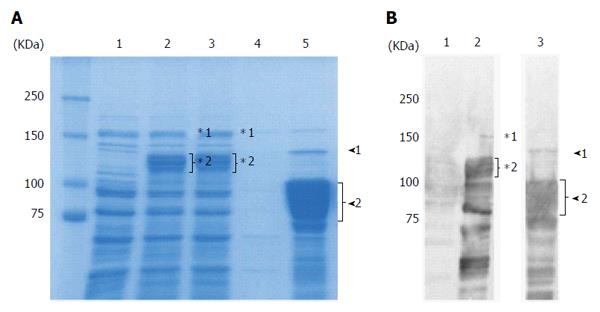
Figure 1 Purification of recombinant East Asian-type CagA.
A: CBB-stained 8% SDS-PAGE gel; 20 μg of protein were loaded per well. Lane 1: Uninduced lysate; 2: 0.4 mmol/L IPTG induced lysate; 3: Flow through; 4: Wash; 5: Elution. In the elution fraction (Lane 5), the 135 kDa band (arrowhead 1) is the full length rCagA. The 75-100 kDa rCagA cleavage product (arrowhead 2) comprised approximately 50% of the elution fraction; B: Western blot analysis with anti-CagA antibody. Lane 1: Uninduced lysate; 2: 0.4 mmol/L IPTG induced lysate; 3: Elution. Following induction (Lane 2), the full length GST-fused rCagA (*1) was expressed. However, it was subsequently cleaved (*2). In the elution fraction (Lane 3), the amount of full length rCagA was low (arrowhead 1) and various smaller-sized bands (arrowhead 2) were confirmed as rCagA fragments.
- Citation: Matsuo Y, Kido Y, Akada J, Shiota S, Binh TT, Trang TTH, Dung HDQ, Tung PH, Tri TD, Thuan NPM, Tam LQ, Nam BC, Khien VV, Yamaoka Y. Novel CagA ELISA exhibits enhanced sensitivity of Helicobacter pylori CagA antibody. World J Gastroenterol 2017; 23(1): 48-59
- URL: https://www.wjgnet.com/1007-9327/full/v23/i1/48.htm
- DOI: https://dx.doi.org/10.3748/wjg.v23.i1.48


