Copyright
©The Author(s) 2016.
World J Gastroenterol. Feb 14, 2016; 22(6): 2142-2148
Published online Feb 14, 2016. doi: 10.3748/wjg.v22.i6.2142
Published online Feb 14, 2016. doi: 10.3748/wjg.v22.i6.2142
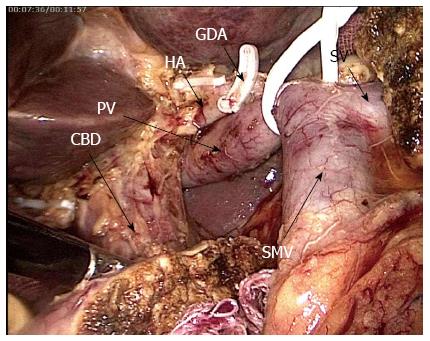
Figure 2 Surgical method - specimen dissection.
After entering the hepatoduodenal ligament, dissecting GDA, and freeing towards the hepatic porta along the surface of hepatic portal vein, it is separated from the hepatic artery and common bile duct. The hepatic artery is fully dissected, and the surrounding fat lymphoid tissues are also dissected. Finally, the common bile duct is dissected. GDA: Gastric-duodenum artery; HA: Hepatic artery; PV: Portal vein; CBD: Common bile duct; SMV: Superior mesenteric vein; SV: Spleen vein.
- Citation: Wang XM, Sun WD, Hu MH, Wang GN, Jiang YQ, Fang XS, Han M. Inferoposterior duodenal approach for laparoscopic pancreaticoduodenectomy. World J Gastroenterol 2016; 22(6): 2142-2148
- URL: https://www.wjgnet.com/1007-9327/full/v22/i6/2142.htm
- DOI: https://dx.doi.org/10.3748/wjg.v22.i6.2142


