Copyright
©The Author(s) 2016.
World J Gastroenterol. Nov 28, 2016; 22(44): 9865-9870
Published online Nov 28, 2016. doi: 10.3748/wjg.v22.i44.9865
Published online Nov 28, 2016. doi: 10.3748/wjg.v22.i44.9865
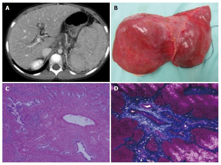
Figure 1 Computed tomography scan (A), macroscopic finding (B) and histology findings (C, D) of the recipient.
A, B: Reveals splenomegaly due to portal hypertension (A), macroscopic finding of the recipient’s native liver (B). Her native liver shows fibrotic changes in the portal area with proliferation of the pseudocholangiolar ducts, which is consistent with congenital hepatic fibrosis (C, D). C: Hematoxylin-eosin staining; D: Azan staining. Magnification × 100.
- Citation: Yamada N, Sanada Y, Katano T, Tashiro M, Hirata Y, Okada N, Ihara Y, Miki A, Sasanuma H, Urahashi T, Sakuma Y, Mizuta K. Pediatric living donor liver transplantation for congenital hepatic fibrosis using a mother’s graft with von Meyenburg complex: A case report. World J Gastroenterol 2016; 22(44): 9865-9870
- URL: https://www.wjgnet.com/1007-9327/full/v22/i44/9865.htm
- DOI: https://dx.doi.org/10.3748/wjg.v22.i44.9865


