Copyright
©The Author(s) 2016.
World J Gastroenterol. Nov 21, 2016; 22(43): 9525-9533
Published online Nov 21, 2016. doi: 10.3748/wjg.v22.i43.9525
Published online Nov 21, 2016. doi: 10.3748/wjg.v22.i43.9525
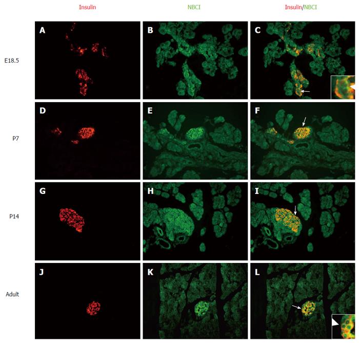
Figure 3 Localization of the NBC1 and insulin determined by immunofluorescence detecting in different developmental stages of rat pancreas.
Labeling of the NBC1 antibodies was detected with an FITC (green)-labeled secondary antibody. Labeling of insulin was detected with a CY3 (red)-labeled secondary antibody on the same section. The overlap of NBC1 (green) and insulin (red) labeling creates orange color. Strong staining for NBC1 is seen in the developing islets at all tested time points, which dominantly colocalizes with insulin positive β cells at the sides of the plasma membrane (arrows). All of the primary magnifications are × 400. Insets, higher magnifications of the areas indicated by arrow in embryonic day (E)18.5 and adult rat. P: Postnatal day.
- Citation: Cao LH, Xia CC, Shi ZC, Wang N, Gu ZH, Yu LZ, Wan Q, De W. Na+/HCO3- cotransporter is expressed on β and α cells during rat pancreatic development. World J Gastroenterol 2016; 22(43): 9525-9533
- URL: https://www.wjgnet.com/1007-9327/full/v22/i43/9525.htm
- DOI: https://dx.doi.org/10.3748/wjg.v22.i43.9525


