Copyright
©The Author(s) 2016.
World J Gastroenterol. Jan 21, 2016; 22(3): 1172-1178
Published online Jan 21, 2016. doi: 10.3748/wjg.v22.i3.1172
Published online Jan 21, 2016. doi: 10.3748/wjg.v22.i3.1172
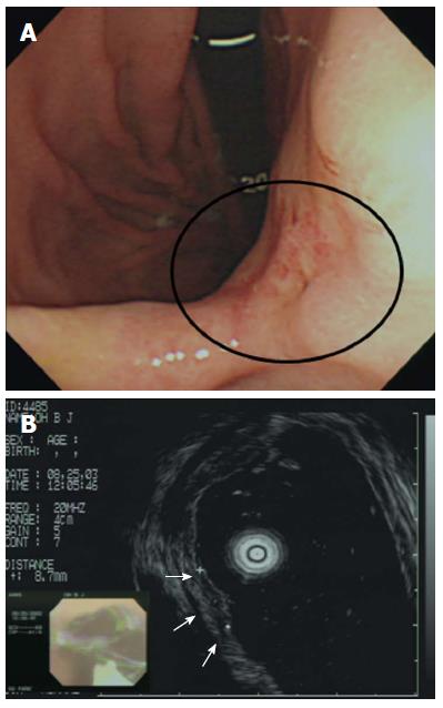
Figure 2 Incorrect T staging: a case of undifferentiated-type early gastric cancer (EGC).
A: Endoscopic image of an EGC showing a 10-mm-diameter depressed lesion in the posterior angle of the wall; B: Endoscopic ultrasonographic image showing a hypoechoic mucosal mass with an intact submucosal layer. A surgical specimen obtained upon radical subtotal gastrectomy confirmed that the EGC was confined to the submucosal layer. Taken with permission from Gastrointest Endosc 2007; 66: 901-908[35].
- Citation: Kim JH. Important considerations when contemplating endoscopic resection of undifferentiated-type early gastric cancer. World J Gastroenterol 2016; 22(3): 1172-1178
- URL: https://www.wjgnet.com/1007-9327/full/v22/i3/1172.htm
- DOI: https://dx.doi.org/10.3748/wjg.v22.i3.1172


