Copyright
©The Author(s) 2016.
World J Gastroenterol. May 14, 2016; 22(18): 4501-4514
Published online May 14, 2016. doi: 10.3748/wjg.v22.i18.4501
Published online May 14, 2016. doi: 10.3748/wjg.v22.i18.4501
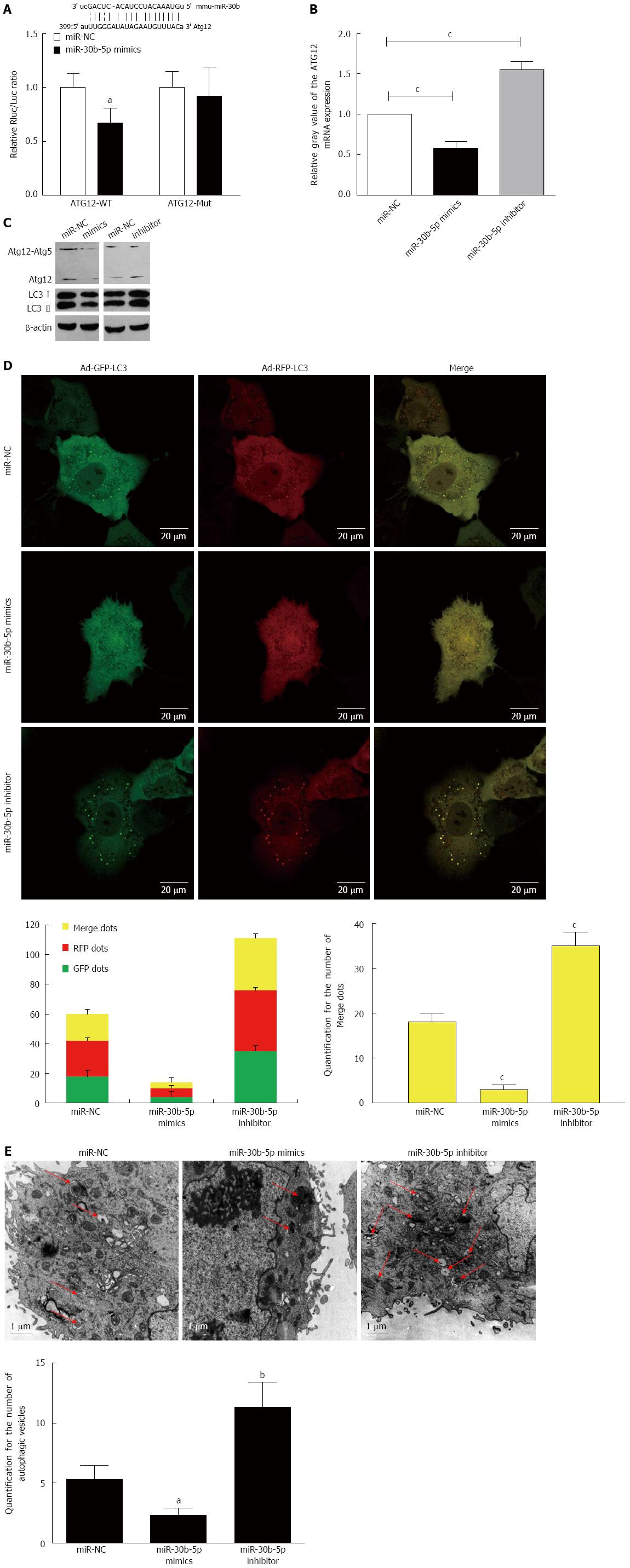
Figure 3 miR-30b inhibits autophagy by sequestering Atg12 in AML12 cells.
A and B: The predicted miR-30b binding site on the Atg12 mRNA 3′-untranslated region (UTR) is shown, and a luciferase reporter assay was performed to determine the effects of miR-30b on the Atg12 mRNA 3′-UTR; C: Western blot was used to detect expression of Atg12 and LC3 in AML12 cells treated with miR-30b mimics or inhibitor; D: Confocal immunofluorescence of AML12 cells demonstrated increased numbers of GFP-RFP-LC3 dots in the IR group; when autophagy is induced, both GFP and RFP are expressed as yellow dots (autophagosomes). Red dots represent autolysosomes as the GFP degrades in an acid environment. Scale bars = 20 μm; E: TEM images of AML12 cells after ischemia followed by reperfusion at 12 h. TEM images show representative examples of autophagosomes (red arrows). Scale bars = 1.0 μm. And the data were quantified by counting the number of autophagosomes per cross-sectioned cell; aP < 0.05, bP < 0.01, cP < 0.001 vs miR-NC group. Every experiment was repeated three times.
- Citation: Li SP, He JD, Wang Z, Yu Y, Fu SY, Zhang HM, Zhang JJ, Shen ZY. miR-30b inhibits autophagy to alleviate hepatic ischemia-reperfusion injury via decreasing the Atg12-Atg5 conjugate. World J Gastroenterol 2016; 22(18): 4501-4514
- URL: https://www.wjgnet.com/1007-9327/full/v22/i18/4501.htm
- DOI: https://dx.doi.org/10.3748/wjg.v22.i18.4501


