Copyright
©The Author(s) 2016.
World J Gastroenterol. Apr 21, 2016; 22(15): 4066-4070
Published online Apr 21, 2016. doi: 10.3748/wjg.v22.i15.4066
Published online Apr 21, 2016. doi: 10.3748/wjg.v22.i15.4066
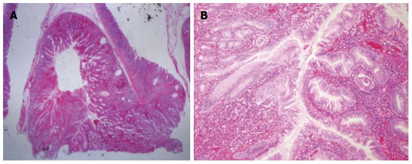
Figure 3 Microscopy of the endoscopic submucosal dissection specimen.
A: Well-circumscribed inverted growth pattern into the submucosal layer was shown (HE staining, × 10); B: Endophytic proliferation of hyperplastic columnar cells and connected with inflamed surface epithelium was shown. No architectural or cytological atypia was found (HE staining, × 100).
- Citation: Yun JT, Lee SW, Kim DP, Choi SH, Kim SH, Park JK, Jang SH, Park YJ, Sung YG, Sul HJ. Gastric inverted hyperplastic polyp: A rare cause of iron deficiency anemia. World J Gastroenterol 2016; 22(15): 4066-4070
- URL: https://www.wjgnet.com/1007-9327/full/v22/i15/4066.htm
- DOI: https://dx.doi.org/10.3748/wjg.v22.i15.4066


