Copyright
©The Author(s) 2016.
World J Gastroenterol. Jan 7, 2016; 22(1): 379-393
Published online Jan 7, 2016. doi: 10.3748/wjg.v22.i1.379
Published online Jan 7, 2016. doi: 10.3748/wjg.v22.i1.379
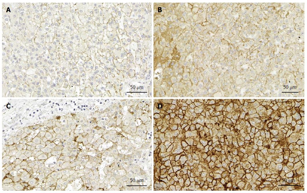
Figure 2 Syndecan expression in the liver.
A: Expression of syndecan-1 in normal liver; B: Expression of syndecan-1 in liver cancer without cirrhosis; C: Expression of syndecan-1 in liver cirrhosis; D: Expression of syndecan-1 in liver cancer with cirrhosis. Note that only modest elevation of syndecan expression can be observed in cancer without cirrhosis, in contrast with the cancer specimen with cirrhosis. Immunopositivity in the cytoplasm of the latter indicates the impairment of transport to the cell surface. Immunohistochemistry on formalin fixed paraffin embedded specimens. (Primary antibody Dako MI 15).
- Citation: Baghy K, Tátrai P, Regős E, Kovalszky I. Proteoglycans in liver cancer. World J Gastroenterol 2016; 22(1): 379-393
- URL: https://www.wjgnet.com/1007-9327/full/v22/i1/379.htm
- DOI: https://dx.doi.org/10.3748/wjg.v22.i1.379


