Copyright
©The Author(s) 2015.
World J Gastroenterol. Feb 14, 2015; 21(6): 1989-1993
Published online Feb 14, 2015. doi: 10.3748/wjg.v21.i6.1989
Published online Feb 14, 2015. doi: 10.3748/wjg.v21.i6.1989
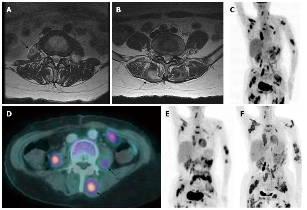
Figure 3 Thoracolumbar spine magnetic resonance imaging and positron-emission tomography-computed tomography images show multiple skeletal metastases in a whole body scan after 3 mo of palliative radiochemotherapy from first diagnosis.
A and B: T2-weighted, axial images show ill-defined, high signal intensity masses in the psoas and paraspinal muscles; C and D: Positron-emission tomography-computed tomography (PET-CT) images show multiple masses with hypermetabolism in the paravertebral area, left upper arm, bilateral periscapular area, anterior and posterior chest walls, bilateral psoas muscles, lower anterior abdominal wall, bilateral gluteal muscles, and both proximal thigh muscles; E and F: Follow-up PET-CT images show aggravated multiple muscle metastasis after 3 and 7 mo of gemcitabine-cisplatin combination chemotherapy.
- Citation: Lee J, Lee SW, Han SY, Baek YH, Kim SY, Rhyou HI. Rapidly aggravated skeletal muscle metastases from an intrahepatic cholangiocarcinoma. World J Gastroenterol 2015; 21(6): 1989-1993
- URL: https://www.wjgnet.com/1007-9327/full/v21/i6/1989.htm
- DOI: https://dx.doi.org/10.3748/wjg.v21.i6.1989


