Copyright
©The Author(s) 2015.
World J Gastroenterol. Feb 14, 2015; 21(6): 1827-1837
Published online Feb 14, 2015. doi: 10.3748/wjg.v21.i6.1827
Published online Feb 14, 2015. doi: 10.3748/wjg.v21.i6.1827
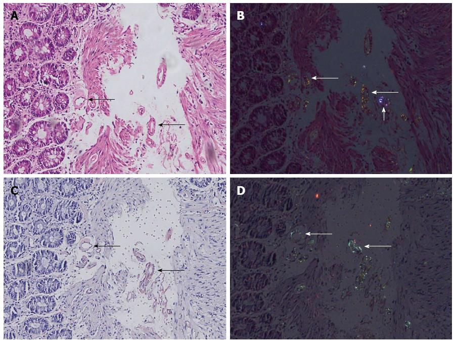
Figure 2 Tiny amyloid deposits in vessel walls, which are not striking at first sight (long arrows).
A: Brightfield view of hematoxylin-eosin (HE) staining; B: Digitally reinforced polarization view of the same HE-stained section. Note the striking yellowish green tint of birefringence of amyloid in small vessel walls (long arrows) and variant colored nonspecific refraction of a foreign body (short arrow); C: Brightfield view of Congo-red staining (note the salmon pink color); D: Polarization view of the same Congo-red-stained section with apple green birefringence of deposits (arrows); 20× magnification.
- Citation: Doganavsargil B, Buberal GE, Toz H, Sarsik B, Pehlivanoglu B, Sezak M, Sen S. Digitally reinforced hematoxylin-eosin polarization technique in diagnosis of rectal amyloidosis. World J Gastroenterol 2015; 21(6): 1827-1837
- URL: https://www.wjgnet.com/1007-9327/full/v21/i6/1827.htm
- DOI: https://dx.doi.org/10.3748/wjg.v21.i6.1827


