Copyright
©The Author(s) 2015.
World J Gastroenterol. Feb 7, 2015; 21(5): 1531-1545
Published online Feb 7, 2015. doi: 10.3748/wjg.v21.i5.1531
Published online Feb 7, 2015. doi: 10.3748/wjg.v21.i5.1531
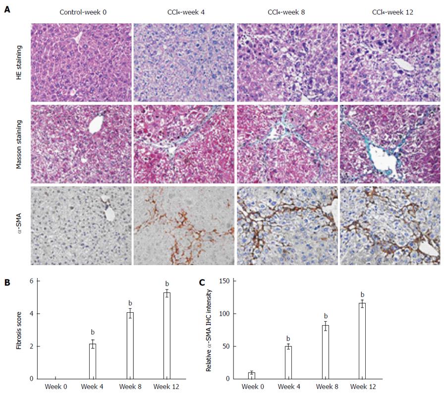
Figure 1 Degree of liver fibrosis in mice was evaluated by histopathological examination.
A: Histology was evaluated by hematoxylin and eosin (HE) staining and fibrillar collagen deposition was assessed by Masson staining (original magnification × 400). Activated hepatic stellate cells were evaluated in liver sections by immunohistochemical staining of alpha-smooth muscle actin (α-SMA) (original magnification × 400); B: Degree of liver fibrosis was assessed based on the Ishak scoring system; C: Morphometric quantitation of α-SMA expression. The mean number of α-SMA-positive cells in five ocular fields (magnification × 400) per specimen was assessed as a percentage area. bP < 0.01 vs the week 0 model group. Data are mean ± SD (n = 8-10).
- Citation: Lu DH, Guo XY, Qin SY, Luo W, Huang XL, Chen M, Wang JX, Ma SJ, Yang XW, Jiang HX. Interleukin-22 ameliorates liver fibrogenesis by attenuating hepatic stellate cell activation and downregulating the levels of inflammatory cytokines. World J Gastroenterol 2015; 21(5): 1531-1545
- URL: https://www.wjgnet.com/1007-9327/full/v21/i5/1531.htm
- DOI: https://dx.doi.org/10.3748/wjg.v21.i5.1531


