Copyright
©The Author(s) 2015.
World J Gastroenterol. Dec 7, 2015; 21(45): 12981-12986
Published online Dec 7, 2015. doi: 10.3748/wjg.v21.i45.12981
Published online Dec 7, 2015. doi: 10.3748/wjg.v21.i45.12981
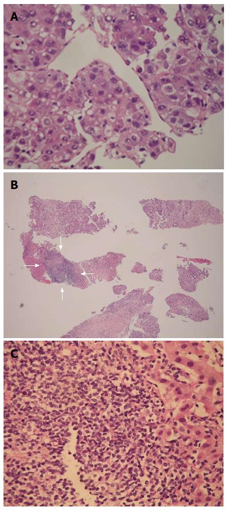
Figure 5 Histological study of the liver tumor.
A: Neoplastic hepatocytes with pleomorphic nuclei and prominent nucleoli; they are considered diagnostic for hepatocellular carcinoma [Hematoxylin and eosin (HE) staining; original magnification × 400]; B: Tissue cores with thick trabeculae of neoplastic hepatocytes and a patchy lymphoid infiltrated (arrows) (HE staining; original magnification × 40); C: Small lymphoid cells with slightly irregular nuclei (HE staining; original magnification × 400).
- Citation: Lee MH, Lin YC, Cheng HT, Chuang WY, Huang HC, Kao HW. Coexistence of hepatoma with mantle cell lymphoma in a hepatitis B carrier. World J Gastroenterol 2015; 21(45): 12981-12986
- URL: https://www.wjgnet.com/1007-9327/full/v21/i45/12981.htm
- DOI: https://dx.doi.org/10.3748/wjg.v21.i45.12981


