Copyright
©The Author(s) 2015.
World J Gastroenterol. Dec 7, 2015; 21(45): 12981-12986
Published online Dec 7, 2015. doi: 10.3748/wjg.v21.i45.12981
Published online Dec 7, 2015. doi: 10.3748/wjg.v21.i45.12981
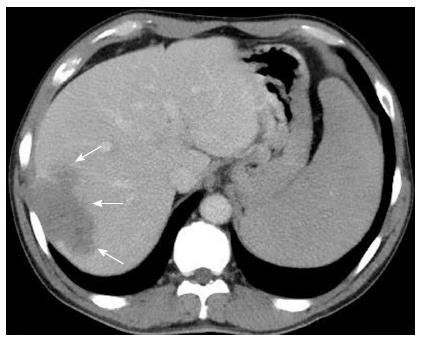
Figure 2 Computed tomography later showed wash out during the delay phase; this is compatible with hepatocellular carcinoma (arrows) with chest wall involvement.
No obvious imaging evidence of liver cirrhosis and portal hypertension was shown.
- Citation: Lee MH, Lin YC, Cheng HT, Chuang WY, Huang HC, Kao HW. Coexistence of hepatoma with mantle cell lymphoma in a hepatitis B carrier. World J Gastroenterol 2015; 21(45): 12981-12986
- URL: https://www.wjgnet.com/1007-9327/full/v21/i45/12981.htm
- DOI: https://dx.doi.org/10.3748/wjg.v21.i45.12981


