Copyright
©The Author(s) 2015.
World J Gastroenterol. Nov 21, 2015; 21(43): 12498-12504
Published online Nov 21, 2015. doi: 10.3748/wjg.v21.i43.12498
Published online Nov 21, 2015. doi: 10.3748/wjg.v21.i43.12498
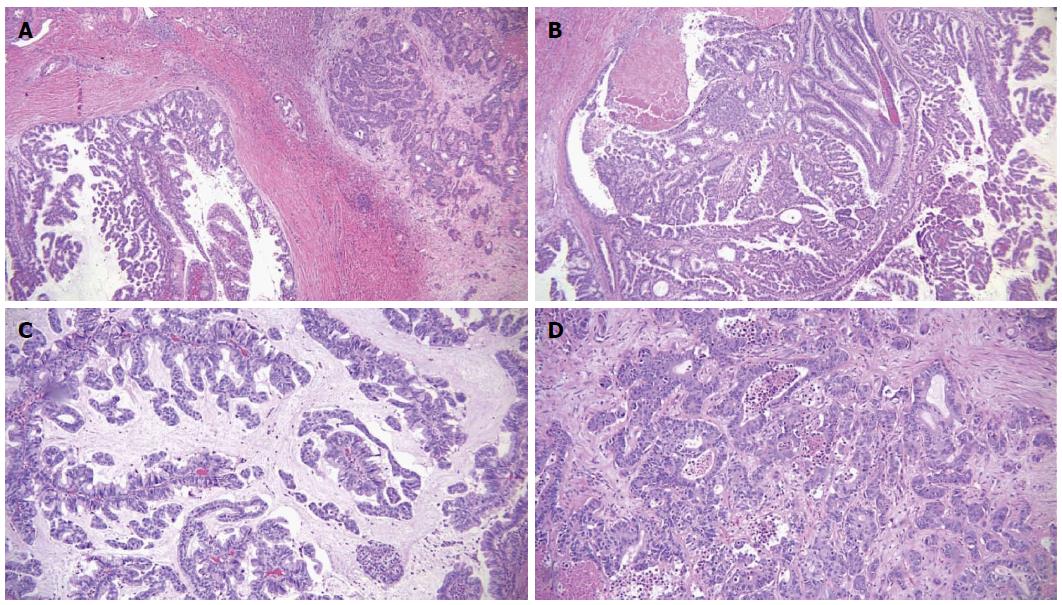
Figure 3 Microscopic features of the tumor.
A: Intraductal papillary mass with adjacent invasive adenocarcinoma; B: The papillary mass with fine vascular cores was lined by foveolar type epithelium; C: Some areas showed mucin production; D: The metastatic masses in the liver showed infiltrative features.
- Citation: Tan Y, Milikowski C, Toribio Y, Singer A, Rojas CP, Garcia-Buitrago MT. Intraductal papillary neoplasm of the bile ducts: A case report and literature review. World J Gastroenterol 2015; 21(43): 12498-12504
- URL: https://www.wjgnet.com/1007-9327/full/v21/i43/12498.htm
- DOI: https://dx.doi.org/10.3748/wjg.v21.i43.12498


