Published online Jan 28, 2015. doi: 10.3748/wjg.v21.i4.1357
Peer-review started: June 6, 2014
First decision: July 21, 2014
Revised: August 6, 2014
Accepted: September 29, 2014
Article in press: September 30, 2014
Published online: January 28, 2015
Carcinosarcoma of the pancreas is an extremely rare tumor and has a dismal prognosis. To the best of our knowledge, the histopathological features of the lesion have been illustrated in the literature but to date no reported cases have been documented on imaging characteristics. We report a female case of pancreatic carcinosarcoma presenting as a mucinous cystadenoma on computed tomography (CT). We also summarize the CT characteristics according to the appended CT images in the reported cases. This is the first report of CT features of pancreatic carcinosarcoma in the English literature.
Core tip: Pancreatic carcinosarcoma is an extremely rare tumor with a poor prognosis. We report a case of pancreatic carcinosarcoma in a 74-year-old woman and describe its appearance on computed tomography (CT). Detailed analysis and conclusion of the CT characteristics were performed according to the appended CT images in reported cases. This is the first report of radiological findings of carcinosarcoma originating from the pancreas.
- Citation: Shi HY, Xie J, Miao F. Pancreatic carcinosarcoma: First literature report on computed tomography imaging. World J Gastroenterol 2015; 21(4): 1357-1361
- URL: https://www.wjgnet.com/1007-9327/full/v21/i4/1357.htm
- DOI: https://dx.doi.org/10.3748/wjg.v21.i4.1357
Pancreatic carcinosarcoma is an uncommon entity comprising a fairly small subset of all pancreatic neoplasms. It is histologically characterized by a mixture of carcinomatous and sarcomatous elements. The prognosis of pancreatic carcinosarcoma is very poor. From the data of reported cases, the majority of patients survived an average of only 6 mo after surgery. Because of the rarity and difficulty of diagnosis, radiological findings of the lesion have not been well illustrated in the literature. Here, we report the clinical and contrast-enhanced computed tomography (CT) findings of carcinosarcoma originating from the pancreas in a 74-year-old woman.
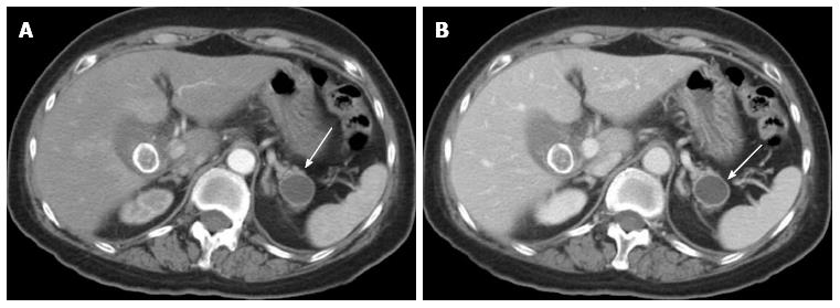
A 74-year-old woman was referred to our hospital with acute calculous cholecystitis. Contrast material-enhanced dual-phase multidetector row CT of the abdomen was performed and revealed a 2.2 cm × 2.0 cm well-circumscribed cystic lesion that was located at the pancreatic tail and did not invade the adjacent viscera. After intravenous injection of contrast media, the lesion had a peripheral enhancing thick wall surrounding the non-enhancing low-attenuation area which is consistent with cystic fluid (Figure 1). Mural nodules and intratumoral septa were not seen on the CT imaging. There was no main pancreatic duct dilatation or any abnormalities in the rest of the pancreatic tissue. We diagnosed it as mucinous cystadenoma. The patient underwent a cholecystectomy and was then discharged.
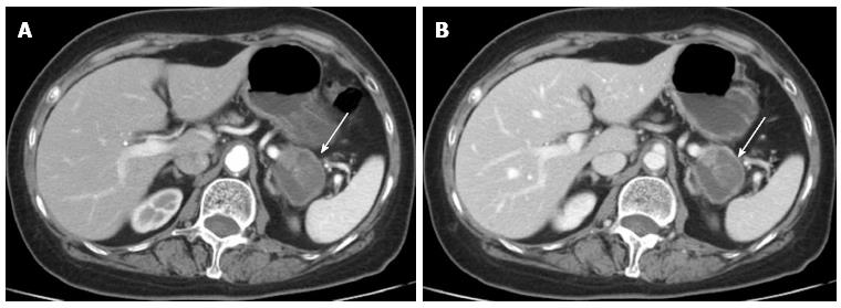
Follow-up CT imaging was performed 13 mo later. The lesion in the pancreatic tail had grown up to 2.9 cm in diameter with a more thickened wall and a solid component appeared in the cystic lesion. The arterial phase revealed heterogeneous enhancement in the wall and solid part of the mass (Figure 2A). In the portal phase, the enhancement became more pronounced. The cystic part revealed non-enhancing low-attenuation (Figure 2B).
After nearly six months, the patient was referred to our hospital again for intermittent abdominal pain and distention lasting for more than 2 wk. She reported no association with food. The abdomen was soft and tender at physical examination. Laboratory analysis revealed elevated CA19-9 (148.40 U/mL), CA72-4 (19.19 U/mL), CA24-2 (51.7 U/mL) and CEA (10.05 ng/mL) levels. Liver function tests and complete blood counts were all within the normal range. CT showed that the original lesion in the pancreatic tail manifested as a heterogeneous complex mass that contained cystic and mixed solid areas and measured 4.0 cm in diameter. The solid components increased and septa in the lesion were aroused. The lesion showed progressive and heterogeneous enhancement (Figure 3). No evidence of metastasis was identified. The CT findings coupled with tumor markers raised the possibility of pancreatic mucinous cystadenocarcinoma. The patient underwent a distal pancreatectomy with splenectomy, followed by uneventful postoperative course.
On gross examination, the resected specimen consisted of a 9 cm × 6 cm × 3 cm segment of the pancreas with a mass in the tail and the spleen. Sectioning through the mass revealed a well-circumscribed tumor consisting of a 5 cm × 4 cm × 2 cm cystic lesion with a thick wall that measured 0.1-0.3 cm. The cystic lesion contained dark-red substances.
Histologically, the tumor of pancreas showed two components separated from each other. The first component was composed of columnar mucin-producing epithelial cells with marked cellular pleomorphism and prominent mitoses, consistent with carcinoma. The second component revealed a sarcomatous growth pattern composed predominantly of highly cellular areas with spindle cells (Figure 4A). Immunohistochemically, the carcinoma component was strongly reactive for antibodies to cytokeratin 7, cytokeratin 19 and cytokeratin AE1/AE3 (Figure 4B). The sarcomatous component was strongly reactive for vimentin (Figure 4C). According to the morphology and the immunohistochemical staining pattern, pancreatic carcinosarcoma was diagnosed.
The concept of carcinosarcoma is that a malignant neoplasm is composed of an intimate admixture of carcinomatous and sarcomatous elements, without areas of transition between both components, and with each of these elements showing distinct immunohistochemical or ultrastructural features pertaining to their different lines of differentiation. Cases fulfilling these criteria have been reported in many organs but rarely in the pancreas. The origin of mixed carcinosarcoma is unknown. Although controversy remains, several studies using diverse immunohistochemical and molecular analyses have suggested that pancreatic carcinosarcoma could be of monoclonal origin and that the sarcomatous component might have arisen from metaplastic transformation of the carcinomatous component[1,2].
The carcinomatous components are varied. Pancreatic ductal adenocarcinoma is the most commonly reported, followed by mucinous cystadenocarcinoma[3-6]. Okamura et al[7] reported intraductal papillary mucinous carcinoma (IPMC) in pancreatic carcinosarcoma for the first time, which is extremely rare. There are also different sarcomatous elements, including spindle cell sarcoma, leiomyosarcoma, malignant fibrous histiocytoma and osteosarcoma.
From the summary of the reported literature, we found that pancreatic carcinosarcoma is common in middle aged and elderly people and few patients were identified incidentally. For patients with symptoms, the most common were abdominal pain, anorexia, nausea and vomiting. When tumors are located in the pancreatic head, they cause early jaundice, commonly seen in other malignant neoplasms. Serum CA19-9 can be elevated in some patients[7-9].
Due to the rarity of this tumor, the prognosis has not been well defined. According to the reported literature, the majority of patients survived an average of only 6 mo after surgery. Zhu et al[9] reported that a 53-year-old woman had remained free of recurrence for 20 mo, the longest recurrence-free survival time recorded for this tumor. Pancreatic carcinosarcoma can also disseminate and recur[10,11]. However, a strategy to improve the prognosis is still not available because of the limited experience of it.
To our best knowledge, the imaging features of pancreatic carcinosarcoma have not been reported. By summarizing the reported cases in the literature, combined with our case, we found that the preferential location for pancreatic carcinosarcoma is the pancreatic head and tail and the size is variable (ranging from 2.5 cm to 20 cm). It is worth noting that pancreatic carcinosarcoma can grow quickly, as observed in our case. During follow-up, the lesion in the pancreatic tail grew from 2.2 cm to 4.0 cm in diameter and a more solid component was also seen 18 mo later. On CT images, the lesion in the pancreatic head more frequently appears as a solid mass with cystic regions and necrosis, while the one in the pancreatic tail is characterized by a cystic neoplasm with mural nodules and a solid component, without accompanying ductal dilatation, although with some exceptions[1,7,11,12]. Tumors located in the pancreatic head can cause pancreatic main duct and intra- and extra-hepatic bile duct dilatation. After intravenous injection of a gadolinium chelate, the solid component, mural nodules and cystic wall show moderate enhancement. Unlike pancreatic ductal adenocarcinoma, carcinosarcoma of the pancreas often has a well-circumscribed border and seldom invades the adjacent organs, extrapancreatic nerve and vascular system. However, they easily metastasize to the liver and peritoneum which is the main cause of death[1,10].
The predominant differential diagnosis with pancreatic carcinosarcoma is pancreatic ductal adenocarcinoma. Compared with ductal adenocarcinoma that is poorly vascularized, pancreatic carcinosarcoma has more vascularity. In addition, extrapancreatic perineural and vascular invasion, atrophy of the pancreatic parenchyma and duct dilatation are less common in carcinosarcoma. When pancreatic carcinosarcoma manifests as a cystic tumor, it is difficult to distinguish it from mucinous cystadenoma or cystadenocarcinoma. They share many features in common, such as focal thickening of the wall, heterogeneous content, mural nodules and so on. However, the preponderance of calcification in the septa and cystic wall enable one to readily distinguish this entity from pancreatic carcinosarcoma because intratumoral calcification in pancreatic carcinosarcoma has not been described so far.
Given the tumor’s rarity, it is difficult to establish more classical imaging findings. Nevertheless, a few characteristic imaging features have been described in this paper. We believe that the characterization of imaging features of pancreatic carcinosarcoma can increase the awareness of this entity among radiologists.
A 74-year-old woman was referred to our hospital for acute calculous cholecystitis and a lesion in the pancreatic tail was discovered. The patient was referred again to our hospital for intermittent abdominal pain and distention lasting for more than 2 wk.
Physical examination showed tenderness on the upper abdominal region.
Pancreatic mucinous cystadenoma or cystadenocarcinoma.
Laboratory analysis revealed elevated CA19-9 (148.40 U/mL), CA72-4 (19.19 U/mL), CA24-2 (51.7 U/mL) and CEA (10.05 ng/mL) levels.
Computed tomography showed that a mass in the pancreatic tail manifested as a heterogeneous complex mass that contained cystic and mixed solid areas and measured 4.0 cm in diameter.
Histological examination demonstrated characteristic histological findings of an intimate admixture of carcinomatous and sarcomatous elements.
The patient underwent a distal pancreatectomy with splenectomy.
The histopathological features of pancreatic carcinosarcoma have been illustrated in the literature but to date no reported cases have been documented on imaging characteristics.
The predominant differential diagnosis of pancreatic carcinosarcoma is pancreatic ductal adenocarcinoma, mucinous cystadenoma or cystadenocarcinoma.
This study determines the histopathological features of carcinosarcoma lesions of the pancreas as this is an extremely rare tumor with a poor prognosis. The authors report the computed tomography (CT) appearance of pancreatic carcinosarcoma which presents as a mucinous cystadenoma in one female patient. Detailed analysis and conclusion of the CT characteristics were performed according to the appended CT images in other reported cases. The authors claim that this is the first report of CT features of pancreatic carcinosarcoma in the English literature.
P- Reviewer: Chowdhury P, Tellez-Avila F S- Editor: Ma YJ L- Editor: Roemmele A E- Editor: Wang CH
| 1. | Kim HS, Joo SH, Yang DM, Lee SH, Choi SH, Lim SJ. Carcinosarcoma of the pancreas: a unique case with emphasis on metaplastic transformation and the presence of undifferentiated pleomorphic high-grade sarcoma. J Gastrointestin Liver Dis. 2011;20:197-200. [PubMed] [Cited in This Article: ] |
| 2. | Wada H, Enomoto T, Fujita M, Yoshino K, Nakashima R, Kurachi H, Haba T, Wakasa K, Shroyer KR, Tsujimoto M. Molecular evidence that most but not all carcinosarcomas of the uterus are combination tumors. Cancer Res. 1997;57:5379-5385. [PubMed] [Cited in This Article: ] |
| 3. | Darvishian F, Sullivan J, Teichberg S, Basham K. Carcinosarcoma of the pancreas: a case report and review of the literature. Arch Pathol Lab Med. 2002;126:1114-1117. [PubMed] [Cited in This Article: ] |
| 4. | Bloomston M, Chanona-Vilchis J, Ellison EC, Ramirez NC, Frankel WL. Carcinosarcoma of the pancreas arising in a mucinous cystic neoplasm. Am Surg. 2006;72:351-355. [PubMed] [Cited in This Article: ] |
| 5. | Barkatullah SA, Deziel DJ, Jakate SM, Kluskens L, Komanduri S. Pancreatic carcinosarcoma with unique triphasic histological pattern. Pancreas. 2005;31:291-292. [PubMed] [DOI] [Cited in This Article: ] [Cited by in Crossref: 16] [Cited by in F6Publishing: 17] [Article Influence: 0.9] [Reference Citation Analysis (0)] |
| 6. | Millis JM, Chang B, Zinner MJ, Barsky SH. Malignant mixed tumor (carcinosarcoma) of the pancreas: a case report supporting organ-induced differentiation of malignancy. Surgery. 1994;115:132-137. [PubMed] [Cited in This Article: ] |
| 7. | Okamura J, Sekine S, Nara S, Ojima H, Shimada K, Kanai Y, Hiraoka N. Intraductal carcinosarcoma with a heterologous mesenchymal component originating in intraductal papillary-mucinous carcinoma (IPMC) of the pancreas with both carcinoma and osteosarcoma cells arising from IPMC cells. J Clin Pathol. 2010;63:266-269. [PubMed] [DOI] [Cited in This Article: ] [Cited by in Crossref: 11] [Cited by in F6Publishing: 11] [Article Influence: 0.8] [Reference Citation Analysis (0)] |
| 8. | Nakano T, Sonobe H, Usui T, Yamanaka K, Ishizuka T, Nishimura E, Hanazaki K. Immunohistochemistry and K-ras sequence of pancreatic carcinosarcoma. Pathol Int. 2008;58:672-677. [PubMed] [DOI] [Cited in This Article: ] [Cited by in Crossref: 22] [Cited by in F6Publishing: 23] [Article Influence: 1.4] [Reference Citation Analysis (0)] |
| 9. | Zhu WY, Liu TG, Zhu H. Long-term recurrence-free survival in a patient with pancreatic carcinosarcoma: a case report with a literature review. Med Oncol. 2012;29:140-143. [PubMed] [DOI] [Cited in This Article: ] [Cited by in Crossref: 20] [Cited by in F6Publishing: 16] [Article Influence: 1.2] [Reference Citation Analysis (0)] |
| 10. | Yamazaki K. A unique pancreatic ductal adenocarcinoma with carcinosarcomatous histology, immunohistochemical distribution of hCG-beta, and the elevation of serum alpha-feto-protein. J Submicrosc Cytol Pathol. 2003;35:343-349. [PubMed] [Cited in This Article: ] |
| 11. | Shen ZL, Wang S, Ye YJ, Wang YL, Sun KK, Yang XD, Jiang KW. Carcinosarcoma of pancreas with liver metastasis combined with gastrointestinal stromal tumour of the stomach: is there a good prognosis with the complete resection? Eur J Cancer Care (Engl). 2010;19:118-123. [PubMed] [DOI] [Cited in This Article: ] [Cited by in Crossref: 25] [Cited by in F6Publishing: 25] [Article Influence: 1.7] [Reference Citation Analysis (0)] |


