Copyright
©The Author(s) 2015.
World J Gastroenterol. Sep 7, 2015; 21(33): 9693-9706
Published online Sep 7, 2015. doi: 10.3748/wjg.v21.i33.9693
Published online Sep 7, 2015. doi: 10.3748/wjg.v21.i33.9693
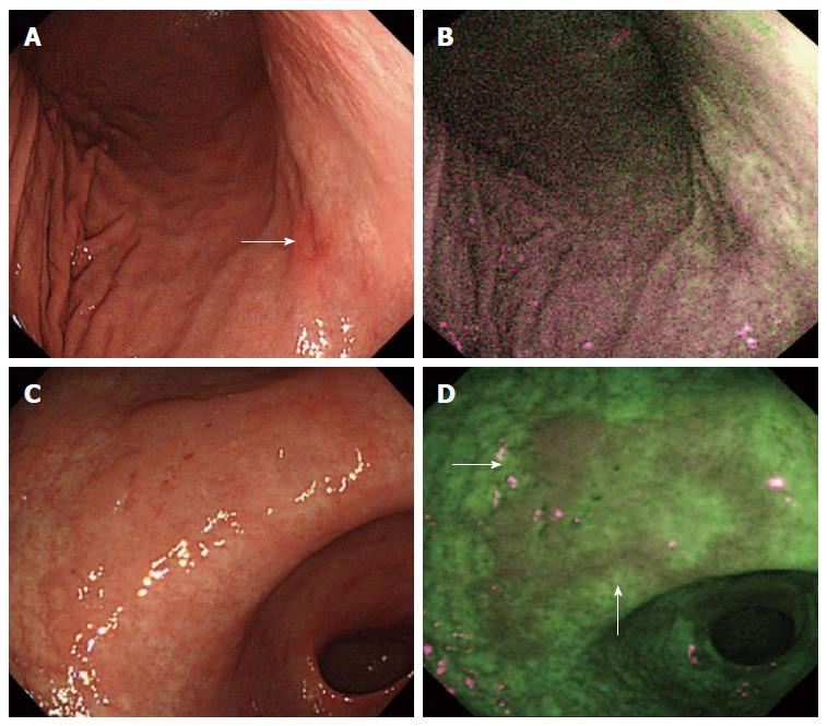
Figure 3 Autofluorescence imaging’s limited role in certain lesions and benefit in others.
A: White light imaging (WLI) displays a red lesion with a slightly depressed area on the bottom right corner (white arrow); B: Autofluorescence imaging (AFI) does not demonstrate a distinct lesion that can be ruled neoplastic; C: WLI made it difficult for the endoscopists to identify the isochromatic flat lesion: D: AFI displays a well-marked lesion with slightly raised borders (white arrows)[50].
- Citation: Ro TH, Mathew MA, Misra S. Value of screening endoscopy in evaluation of esophageal, gastric and colon cancers. World J Gastroenterol 2015; 21(33): 9693-9706
- URL: https://www.wjgnet.com/1007-9327/full/v21/i33/9693.htm
- DOI: https://dx.doi.org/10.3748/wjg.v21.i33.9693


