Copyright
©The Author(s) 2015.
World J Gastroenterol. Jan 21, 2015; 21(3): 759-785
Published online Jan 21, 2015. doi: 10.3748/wjg.v21.i3.759
Published online Jan 21, 2015. doi: 10.3748/wjg.v21.i3.759
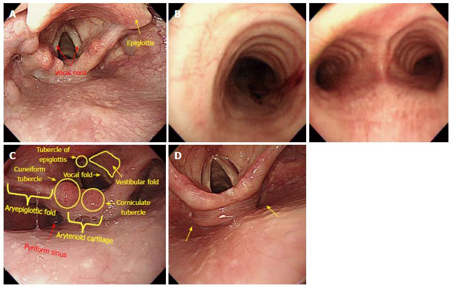
Figure 13 Intubation from hypopharynx to upper esophagus.
A: Hypopharynx. The hypopharynx refers to a part of the pharynx located in the posterior of the larynx (usually from the epiglottis to the vocal cord); B: Bronchus. If the scope is mistakenly inserted into the vocal cord, the circular cricoid cartilage, which belongs to the main bronchus (left), is observed. If the scope is passed further without noticing that it has been placed in a wrong position, the scope will enter the area (right) where it bifurcates into the left and right sides. When an examinee suffers a coughing reflex together with severe dyspnea, the scope must be immediately withdrawn; C: Anatomical structures of hypopharynx and larynx; D: Left- and right-sided pyriform fossa. As an examinee lies in a left lateral decubitus position, it is usually recommended to insert the scope into the left sided pyriform fossa. However, after several attempts to insert the scope have failed and ended with damaged tissues, advancement into the right-sided pyriform fossa should be attempted.
- Citation: Lee SH, Park YK, Cho SM, Kang JK, Lee DJ. Technical skills and training of upper gastrointestinal endoscopy for new beginners. World J Gastroenterol 2015; 21(3): 759-785
- URL: https://www.wjgnet.com/1007-9327/full/v21/i3/759.htm
- DOI: https://dx.doi.org/10.3748/wjg.v21.i3.759


