Copyright
©The Author(s) 2015.
World J Gastroenterol. Jan 21, 2015; 21(3): 759-785
Published online Jan 21, 2015. doi: 10.3748/wjg.v21.i3.759
Published online Jan 21, 2015. doi: 10.3748/wjg.v21.i3.759
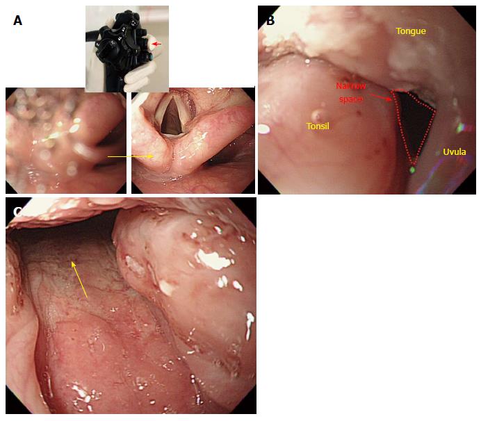
Figure 12 Intubation of scope from oral cavity into pharynx.
A: If the visibility is compromised by saliva in the process of advancement after inserting the scope into the oral cavity (left), one can secure clear visualization of the entrance route (right) by suctioning the saliva with a push on the suction button (small arrow mark). If one advances the scope into the esophagus without suctioning the saliva, an examinee may inhale saliva, which can cause a severe coughing reflex. Therefore, it is recommended to suction saliva during the advancement process; B: Swollen tonsil. As the pathway where the tongue, uvula and tonsil are located together is narrow due to a swollen tonsil, it can be difficult to advance the scope, and a beginner may feel frustrated. However, as the endoscope is ductile, the scope can pass into the oropharynx if it is twisted carefully (to the right, in this case); C: Passage through the swollen tonsil.
- Citation: Lee SH, Park YK, Cho SM, Kang JK, Lee DJ. Technical skills and training of upper gastrointestinal endoscopy for new beginners. World J Gastroenterol 2015; 21(3): 759-785
- URL: https://www.wjgnet.com/1007-9327/full/v21/i3/759.htm
- DOI: https://dx.doi.org/10.3748/wjg.v21.i3.759


