Copyright
©The Author(s) 2015.
World J Gastroenterol. Jan 21, 2015; 21(3): 759-785
Published online Jan 21, 2015. doi: 10.3748/wjg.v21.i3.759
Published online Jan 21, 2015. doi: 10.3748/wjg.v21.i3.759
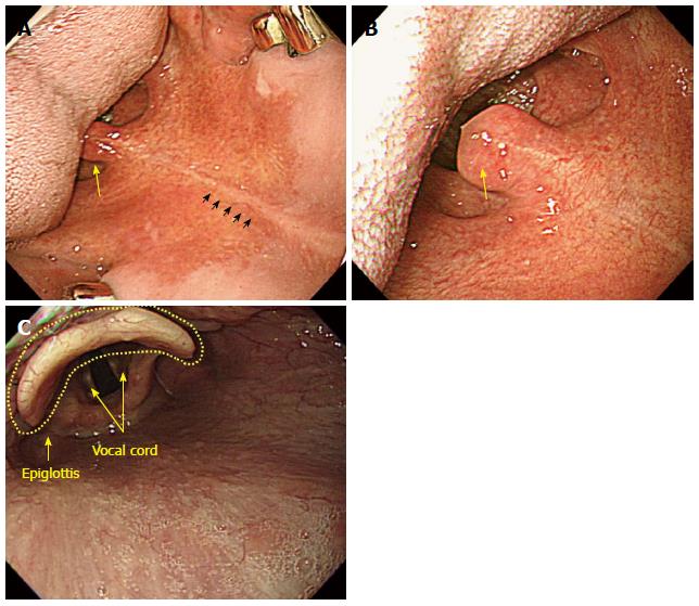
Figure 10 Intubation of scope from oral cavity into pharynx.
A: After placing the tongue in a right position on the screen, an examiner has to advance the scope along the middle line (small arrow marks) of the soft palate. The uvula is observed in the upper side of the tongue (lower side); B: The uvula is observed more clearly at the end of the tongue (tongue base). After that, rotate the scope a little bit to the left and advance it further; C: If the scope is advanced further, it will enter the hypopharynx where the epiglottis is observed together with the vocal cord.
- Citation: Lee SH, Park YK, Cho SM, Kang JK, Lee DJ. Technical skills and training of upper gastrointestinal endoscopy for new beginners. World J Gastroenterol 2015; 21(3): 759-785
- URL: https://www.wjgnet.com/1007-9327/full/v21/i3/759.htm
- DOI: https://dx.doi.org/10.3748/wjg.v21.i3.759


