Copyright
©The Author(s) 2015.
World J Gastroenterol. Jan 21, 2015; 21(3): 759-785
Published online Jan 21, 2015. doi: 10.3748/wjg.v21.i3.759
Published online Jan 21, 2015. doi: 10.3748/wjg.v21.i3.759
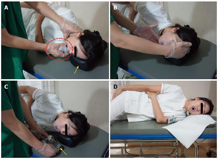
Figure 7 Intubation of scope from oral cavity into pharynx.
A: The examiner is inserting a mouthpiece into the mouth of the examinee (dotted circle). The pillow (arrow mark) helps keep the neck and head straight with the trunk; B: To ensure a smooth insertion of the endoscope, the examiner needs to raise the chin of the examinee and have the head protrude a little bit forward. If this happens, the tongue base separates from the hypopharynx so that the insertion of the endoscope is easier; C: A small container is placed close to the mouth of a patient to collect saliva during and after the esophagogastroduodenoscopy; D: The examinee with a mouthpiece placed in the mouth is fully ready for the examination.
- Citation: Lee SH, Park YK, Cho SM, Kang JK, Lee DJ. Technical skills and training of upper gastrointestinal endoscopy for new beginners. World J Gastroenterol 2015; 21(3): 759-785
- URL: https://www.wjgnet.com/1007-9327/full/v21/i3/759.htm
- DOI: https://dx.doi.org/10.3748/wjg.v21.i3.759


