
















©The Author(s) 2015.
World J Gastroenterol. May 21, 2015; 21(19): 6060-6064
Published online May 21, 2015. doi: 10.3748/wjg.v21.i19.6060
Published online May 21, 2015. doi: 10.3748/wjg.v21.i19.6060
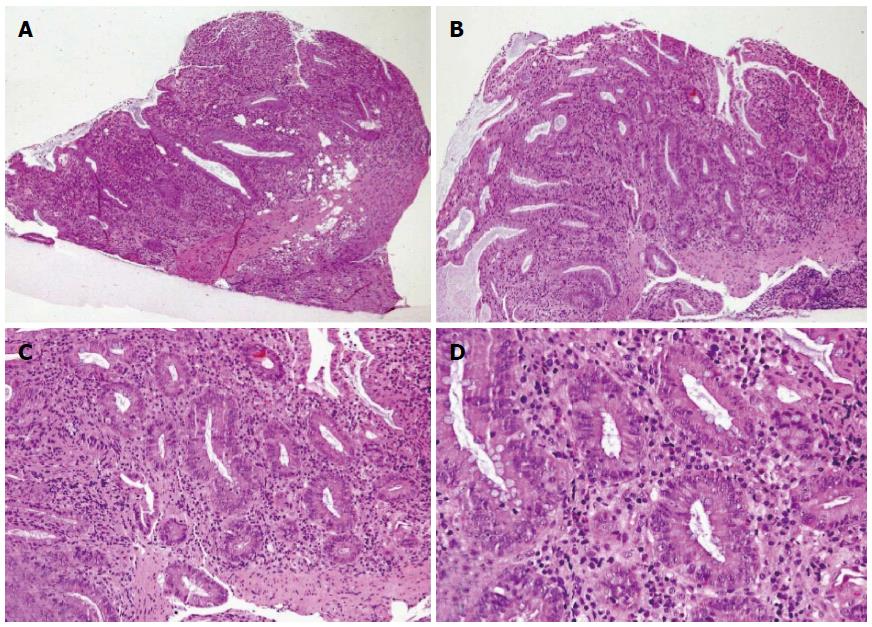
Figure 3 Histopathology of active ulcerative colitis.
Colonic mucosa characterized by atrophic glands with architectural distortion, mucin depletion, intense inflammatory cell infiltrate of the lamina propria rich in eosinophils, basal plasma cells, crypt abscess, and surface ulceration (A, B: Hematoxylin-eosin staining, magnification × 4; C: Hematoxylin-eosin staining, magnification × 10; D: Hematoxylin-eosin staining, magnification × 20).
- Citation: Orabona R, Valcamonico A, Salemme M, Manenti S, Tiberio GA, Frusca T. Fulminant ulcerative colitis in a healthy pregnant woman. World J Gastroenterol 2015; 21(19): 6060-6064
- URL: https://www.wjgnet.com/1007-9327/full/v21/i19/6060.htm
- DOI: https://dx.doi.org/10.3748/wjg.v21.i19.6060