Copyright
©The Author(s) 2015.
World J Gastroenterol. Apr 21, 2015; 21(15): 4583-4591
Published online Apr 21, 2015. doi: 10.3748/wjg.v21.i15.4583
Published online Apr 21, 2015. doi: 10.3748/wjg.v21.i15.4583
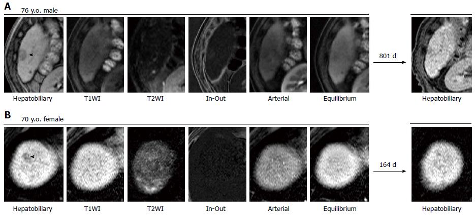
Figure 2 Representative gadolinium ethoxybenzyl diethylene-triamine-pentaacetic-acid magnetic resonance imaging of non-hypervascular nodules that resolved over time.
A: A non-hypervascular nodules (NHNs) was clearly detected at the surface of segment 6 in the hepatobiliary phase, as indicated by an arrowhead in the area ventrolateral to a tiny cyst. The NHN was also revealed in the arterial and equilibrium phases as a lower-intensity nodule, while neither T1-weighted imaging (T1WI) nor T2-weighted imaging visualized the NHN. A fat deposition was expected on the basis of higher intensity in the subtraction of in- and out-of-phase (In-Out) fat-suppressed T1WI. After more than 2 years, the NHN was still there, but the diameter was reduced to less than 5 mm; B: The hepatobiliary phase presented a 6-mm NHN at segment 8 in the vicinity of the diaphragmatic surface of the liver, as indicated by an arrowhead. The NHN could not be visualized in any other sequence. A subsequent gadolinium ethoxybenzyl diethylene-triamine-pentaacetic-acid magnetic resonance imaging showed no nodular lesion in the same area after approximately 5 mo. This NHN was detected together with the other NHN that is presented in Figure 1B.
- Citation: Kanefuji T, Takano T, Suda T, Akazawa K, Yokoo T, Kamimura H, Kamimura K, Tsuchiya A, Takamura M, Kawai H, Yamagiwa S, Aoyama H, Nomoto M, Terai S. Factors predicting aggressiveness of non-hypervascular hepatic nodules detected on hepatobiliary phase of gadolinium ethoxybenzyl diethylene-triamine-pentaacetic-acid magnetic resonance imaging. World J Gastroenterol 2015; 21(15): 4583-4591
- URL: https://www.wjgnet.com/1007-9327/full/v21/i15/4583.htm
- DOI: https://dx.doi.org/10.3748/wjg.v21.i15.4583


