Copyright
©The Author(s) 2015.
World J Gastroenterol. Mar 21, 2015; 21(11): 3409-3413
Published online Mar 21, 2015. doi: 10.3748/wjg.v21.i11.3409
Published online Mar 21, 2015. doi: 10.3748/wjg.v21.i11.3409
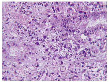
Figure 3 Histologic analysis of the specimen by the endoscopic gastric mucosal biopsy shows round-oval neoplastic cells arranged in abortive glandular pattern or individually over the lamina propria and signet-ring tumor cells are identified.
Hematoxylin and eosin staining, magnification × 400.
- Citation: Lin CH, Lin WC, Lai IH, Wu SF, Wu KH, Chen AC. Pediatric gastric cancer presenting with massive ascites. World J Gastroenterol 2015; 21(11): 3409-3413
- URL: https://www.wjgnet.com/1007-9327/full/v21/i11/3409.htm
- DOI: https://dx.doi.org/10.3748/wjg.v21.i11.3409


