Copyright
©The Author(s) 2015.
World J Gastroenterol. Mar 21, 2015; 21(11): 3394-3401
Published online Mar 21, 2015. doi: 10.3748/wjg.v21.i11.3394
Published online Mar 21, 2015. doi: 10.3748/wjg.v21.i11.3394
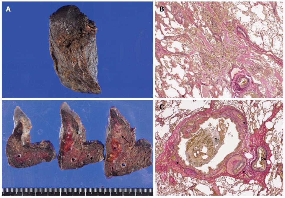
Figure 5 Gross and histopathological findings of the resected specimen.
A: An ill-demarcated whitish area with a brownish tint was noted on the cut surface of the resected lung (arrows). Macroscopically, the bleeding origin was not detected; B: Organizing pneumonia with the accumulation of hemosiderin-laden macrophages was observed [Elastica van Gieson stain (EVG), magnification × 24]; C: The bronchus was enlarged, and engorged bronchial artery with medial hypertrophy and overgrowth of small branches were identified near the bronchus (arrows) (EVG, magnification × 24). Br: Bronchus; PA: Pulmonary artery.
- Citation: Kitajima T, Momose K, Lee S, Haruta S, Ueno M, Shinohara H, Fujimori S, Fujii T, Takei R, Kohno T, Udagawa H. Bronchial bleeding caused by recurrent pneumonia after radical esophagectomy for esophageal cancer. World J Gastroenterol 2015; 21(11): 3394-3401
- URL: https://www.wjgnet.com/1007-9327/full/v21/i11/3394.htm
- DOI: https://dx.doi.org/10.3748/wjg.v21.i11.3394


